6K97

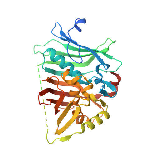
Crystal structure of fusion DH domain


Experimental Data Snapshot

  • Method: X-RAY DIFFRACTION
  • Resolution: 2.50 Å
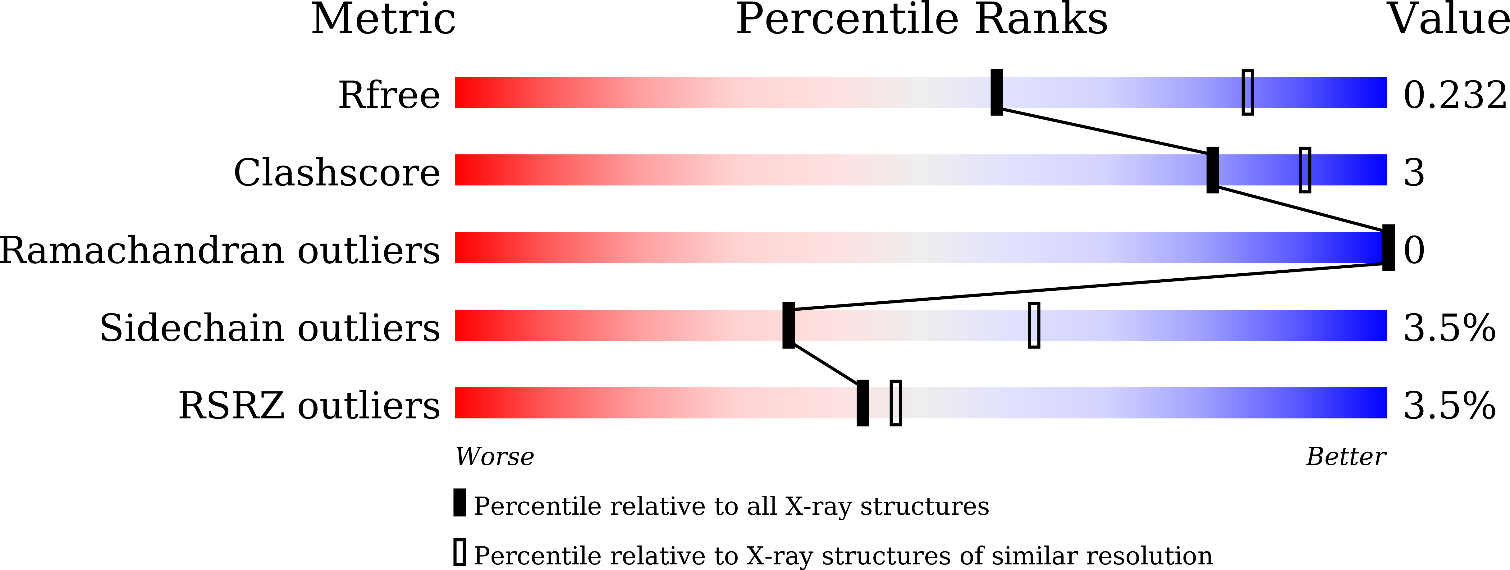
  • R-Value Free: 0.235 
  • R-Value Work: 0.195 
  • R-Value Observed: 0.197 

Starting Model: experimental
View more details

wwPDB Validation   3D Report Full Report


This is version 1.2 of the entry. See complete history


Literature

Functional and Structural Analyses of the Split-Dehydratase Domain in the Biosynthesis of Macrolactam Polyketide Cremimycin.

Kawasaki, D.Miyanaga, A.Chisuga, T.Kudo, F.Eguchi, T.

(2019) Biochemistry 58: 4799-4803

  • DOI: https://doi.org/10.1021/acs.biochem.9b00897
  • Primary Citation of Related Structures:  
    6K97

  • PubMed Abstract: 

    In the biosynthesis of the macrolactam antibiotic cremimycin, the 3-aminononanoic acid starter unit is formed via a non-2-enoyl acyl carrier protein thioester intermediate, which is presumed to be constructed by cis -acyltransferase (AT) polyketide synthases (PKSs) CmiP2, CmiP3, and CmiP4. While canonical cis -AT PKS modules are comprised of a single polypeptide, the PKS module formed by CmiP2 and CmiP3 is split within the dehydratase (DH) domain. Here, we report the enzymatic function and the structural features of this split-DH domain. In vitro analysis showed that the split-DH domain catalyzes the dehydration reaction of ( R )-3-hydroxynonanoyl N -acetylcysteamine thioester (SNAC) to form ( E )-non-2-enoyl-SNAC, suggesting that the split-DH domain is catalytically active in cremimycin biosynthesis. In addition, structural analysis revealed that the CmiP2 and CmiP3 subunits of the split-DH domain form a tightly associated heterodimer through several hydrogen bonding and hydrophobic interactions, which are similar to those of canonical DH domains of other cis -AT PKSs. These results indicate that the split-DH domain has the same function and structure as common cis -AT PKS DH domains.


  • Organizational Affiliation

    Department of Chemistry , Tokyo Institute of Technology , 2-12-1 O-okayama , Meguro-ku, Tokyo 152-8551 , Japan.


Macromolecules
Find similar proteins by:  (by identity cutoff)  |  3D Structure
Entity ID: 1
MoleculeChains Sequence LengthOrganismDetailsImage
Fusion DH
A, B
340Streptomyces sp. MJ635-86F5Mutation(s): 0 
Gene Names: cmiP3cmiP2
UniProt
Find proteins for X5IY86 (Streptomyces sp. MJ635-86F5)
Explore X5IY86 
Go to UniProtKB:  X5IY86
Find proteins for X5IJ93 (Streptomyces sp. MJ635-86F5)
Explore X5IJ93 
Go to UniProtKB:  X5IJ93
Entity Groups  
Sequence Clusters30% Identity50% Identity70% Identity90% Identity95% Identity100% Identity
UniProt GroupsX5IY86X5IJ93
Sequence Annotations
Expand
  • Reference Sequence
Small Molecules
Ligands 1 Unique
IDChains Name / Formula / InChI Key2D Diagram3D Interactions
SO4
Query on SO4

Download Ideal Coordinates CCD File 
C [auth A]SULFATE ION
O4 S
QAOWNCQODCNURD-UHFFFAOYSA-L
Experimental Data & Validation

Experimental Data

  • Method: X-RAY DIFFRACTION
  • Resolution: 2.50 Å
  • R-Value Free: 0.235 
  • R-Value Work: 0.195 
  • R-Value Observed: 0.197 
  • Space Group: C 1 2 1
Unit Cell:
Length ( Å )Angle ( ˚ )
a = 154.632α = 90
b = 66.564β = 90.97
c = 63.013γ = 90
Software Package:
Software NamePurpose
REFMACrefinement
XDSdata reduction
Aimlessdata scaling
MOLREPphasing

Structure Validation

View Full Validation Report


Entry History 

Deposition Data

Revision History  (Full details and data files)

  • Version 1.0: 2019-11-27
    Type: Initial release
  • Version 1.1: 2019-12-11
    Changes: Database references
  • Version 1.2: 2023-11-22
    Changes: Data collection, Database references, Refinement description