6FD3

Thiophosphorylated PAK3 kinase domain


Experimental Data Snapshot

  • Method: X-RAY DIFFRACTION
  • Resolution: 1.52 Å
  • R-Value Free: 0.190 
  • R-Value Work: 0.175 
  • R-Value Observed: 0.176 

Starting Model: experimental
View more details

wwPDB Validation   3D Report Full Report


Ligand Structure Quality Assessment 


This is version 1.2 of the entry. See complete history


Literature

Solution structures and biophysical analysis of full-length group A PAKs reveal they are monomeric and auto-inhibited incis.

Sorrell, F.J.Kilian, L.M.Elkins, J.M.

(2019) Biochem J 476: 1037-1051

  • DOI: https://doi.org/10.1042/BCJ20180867
  • Primary Citation of Related Structures:  
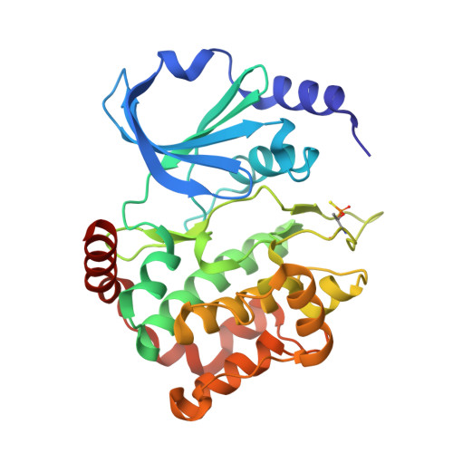
    6FD3

  • PubMed Abstract: 

    The group A p21-activated kinases (PAKs) exist in an auto-inhibited form until activated by GTPase binding and auto-phosphorylation. In the auto-inhibited form, a regulatory domain binds to the kinase domain (KD) blocking the binding of substrates, and CDC42 or Rac binding to the regulatory domain relieves this auto-inhibition allowing auto-phosphorylation on the KD activation loop. We have determined the crystal structure of the PAK3 catalytic domain and by small angle X-ray scattering, the solution-phase structures of full-length inactive PAK1 and PAK3. The structures reveal a compact but elongated molecular shape that demonstrates that, together with multiple independent biophysical measurements and in contrast with previous assumptions, group A PAKs are monomeric both before and after activation, consistent with an activation mechanism of cis -auto-inhibition and initial cis -auto-phosphorylation, followed by transient dimerisation to allow trans -auto-phosphorylation for full activation, yielding a monomeric active PAK protein.


  • Organizational Affiliation

    Structural Genomics Consortium, University of Oxford, Old Road Campus Research Building, Roosevelt Drive, Oxford OX3 7DQ, U.K.


Macromolecules
Find similar proteins by:  (by identity cutoff)  |  3D Structure
Entity ID: 1
MoleculeChains Sequence LengthOrganismDetailsImage
Serine/threonine-protein kinase PAK 3300Homo sapiensMutation(s): 1 
Gene Names: PAK3OPHN3
EC: 2.7.11.1
UniProt & NIH Common Fund Data Resources
Find proteins for O75914 (Homo sapiens)
Explore O75914 
Go to UniProtKB:  O75914
PHAROS:  O75914
GTEx:  ENSG00000077264 
Entity Groups  
Sequence Clusters30% Identity50% Identity70% Identity90% Identity95% Identity100% Identity
UniProt GroupO75914
Sequence Annotations
Expand
  • Reference Sequence
Experimental Data & Validation

Experimental Data

  • Method: X-RAY DIFFRACTION
  • Resolution: 1.52 Å
  • R-Value Free: 0.190 
  • R-Value Work: 0.175 
  • R-Value Observed: 0.176 
  • Space Group: I 41
Unit Cell:
Length ( Å )Angle ( ˚ )
a = 108.825α = 90
b = 108.825β = 90
c = 57.934γ = 90
Software Package:
Software NamePurpose
PHENIXrefinement
XDSdata reduction
Aimlessdata scaling
PHASERphasing
PDB_EXTRACTdata extraction

Structure Validation

View Full Validation Report


Ligand Structure Quality Assessment 


Entry History 

Deposition Data

Revision History  (Full details and data files)

  • Version 1.0: 2018-01-03
    Type: Initial release
  • Version 1.1: 2019-07-17
    Changes: Data collection, Database references
  • Version 1.2: 2024-01-17
    Changes: Data collection, Database references, Derived calculations, Refinement description