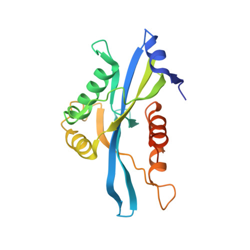
6B09

Crystal structure of HsNUDT16 in complex with diADPR (soaked)


Experimental Data Snapshot

  • Method: X-RAY DIFFRACTION
  • Resolution: 3.20 Å
  • R-Value Free: 0.279 
  • R-Value Work: 0.256 
  • R-Value Observed: 0.257 

wwPDB Validation   3D Report Full Report


Ligand Structure Quality Assessment 


This is version 1.2 of the entry. See complete history


Literature

Structural analyses of NudT16-ADP-ribose complexes direct rational design of mutants with improved processing of poly(ADP-ribosyl)ated proteins.

Thirawatananond, P.McPherson, R.L.Malhi, J.Nathan, S.Lambrecht, M.J.Brichacek, M.Hergenrother, P.J.Leung, A.K.L.Gabelli, S.B.

(2019) Sci Rep 9: 5940-5940

  • DOI: https://doi.org/10.1038/s41598-019-39491-w
  • Primary Citation of Related Structures:  
    5VY2, 5W6X, 5W6Z, 5WJI, 6B09

  • PubMed Abstract: 

    ADP-ribosylation is a post-translational modification that occurs on chemically diverse amino acids, including aspartate, glutamate, lysine, arginine, serine and cysteine on proteins and is mediated by ADP-ribosyltransferases, including a subset commonly known as poly(ADP-ribose) polymerases. ADP-ribose can be conjugated to proteins singly as a monomer or in polymeric chains as poly(ADP-ribose). While ADP-ribosylation can be reversed by ADP-ribosylhydrolases, this protein modification can also be processed to phosphoribosylation by enzymes possessing phosphodiesterase activity, such as snake venom phosphodiesterase, mammalian ectonucleotide pyrophosphatase/phosphodiesterase 1, Escherichia coli RppH, Legionella pneumophila Sde and Homo sapiens NudT16 (HsNudT16). Our studies here sought to utilize X-ray crystallographic structures of HsNudT16 in complex with monomeric and dimeric ADP-ribose in identifying the active site for binding and processing free and protein-conjugated ADP-ribose into phosphoribose forms. These structural data guide rational design of mutants that widen the active site to better accommodate protein-conjugated ADP-ribose. We identified that several HsNudT16 mutants (Δ17, F36A, and F61S) have reduced activity for free ADP-ribose, similar processing ability against protein-conjugated mono(ADP-ribose), but improved catalytic efficiency for protein-conjugated poly(ADP-ribose). These HsNudT16 variants may, therefore, provide a novel tool to investigate different forms of ADP-ribose.


  • Organizational Affiliation

    Department of Biophysics and Biophysical Chemistry, Johns Hopkins University School of Medicine, Baltimore, MD, 21205, USA.


Macromolecules
Find similar proteins by:  (by identity cutoff)  |  3D Structure
Entity ID: 1
MoleculeChains Sequence LengthOrganismDetailsImage
U8 snoRNA-decapping enzyme
A, B
195Homo sapiensMutation(s): 1 
Gene Names: NUDT16
EC: 3.6.1.62 (PDB Primary Data), 3.6.1.64 (PDB Primary Data)
UniProt & NIH Common Fund Data Resources
Find proteins for Q96DE0 (Homo sapiens)
Explore Q96DE0 
Go to UniProtKB:  Q96DE0
PHAROS:  Q96DE0
GTEx:  ENSG00000198585 
Entity Groups  
Sequence Clusters30% Identity50% Identity70% Identity90% Identity95% Identity100% Identity
UniProt GroupQ96DE0
Sequence Annotations
Expand
  • Reference Sequence
Small Molecules
Ligands 4 Unique
IDChains Name / Formula / InChI Key2D Diagram3D Interactions
C7G
Query on C7G

Download Ideal Coordinates CCD File 
C [auth A],
H [auth B]
[(2~{R},3~{S},4~{S},5~{S})-5-(6-aminopurin-9-yl)-4-[(2~{S},3~{S},4~{S},5~{S})-5-[[[[(2~{R},3~{R},4~{S},5~{S})-5-(6-aminopurin-9-yl)-3,4-bis(oxidanyl)oxolan-2-yl]methoxy-oxidanyl-phosphoryl]oxy-oxidanyl-phosphoryl]oxymethyl]-3,4-bis(oxidanyl)oxolan-2-yl]oxy-3-oxidanyl-oxolan-2-yl]methyl phosphono hydrogen phosphate
C25 H36 N10 O23 P4
CSEMRHUJDKFXPV-OATIKAJMSA-N
CL
Query on CL

Download Ideal Coordinates CCD File 
F [auth A]CHLORIDE ION
Cl
VEXZGXHMUGYJMC-UHFFFAOYSA-M
MG
Query on MG

Download Ideal Coordinates CCD File 
D [auth A],
E [auth A],
I [auth B],
J [auth B]
MAGNESIUM ION
Mg
JLVVSXFLKOJNIY-UHFFFAOYSA-N
NA
Query on NA

Download Ideal Coordinates CCD File 
G [auth A],
K [auth B]
SODIUM ION
Na
FKNQFGJONOIPTF-UHFFFAOYSA-N
Experimental Data & Validation

Experimental Data

  • Method: X-RAY DIFFRACTION
  • Resolution: 3.20 Å
  • R-Value Free: 0.279 
  • R-Value Work: 0.256 
  • R-Value Observed: 0.257 
  • Space Group: C 1 2 1
Unit Cell:
Length ( Å )Angle ( ˚ )
a = 113.395α = 90
b = 47.063β = 108.74
c = 75.792γ = 90
Software Package:
Software NamePurpose
REFMACrefinement
StructureStudiodata collection
SCALEPACKdata scaling
PDB_EXTRACTdata extraction
Cootmodel building
HKL-3000data reduction
MOLREPphasing

Structure Validation

View Full Validation Report


Ligand Structure Quality Assessment 


Entry History 

Deposition Data

Revision History  (Full details and data files)

  • Version 1.0: 2019-01-09
    Type: Initial release
  • Version 1.1: 2019-04-24
    Changes: Data collection, Database references
  • Version 1.2: 2024-03-13
    Changes: Data collection, Database references, Derived calculations