6AC3

Structure of a natural red emitting luciferase from Phrixothrix hirtus (P3121 crystal form)


Experimental Data Snapshot

  • Method: X-RAY DIFFRACTION
  • Resolution: 3.60 Å
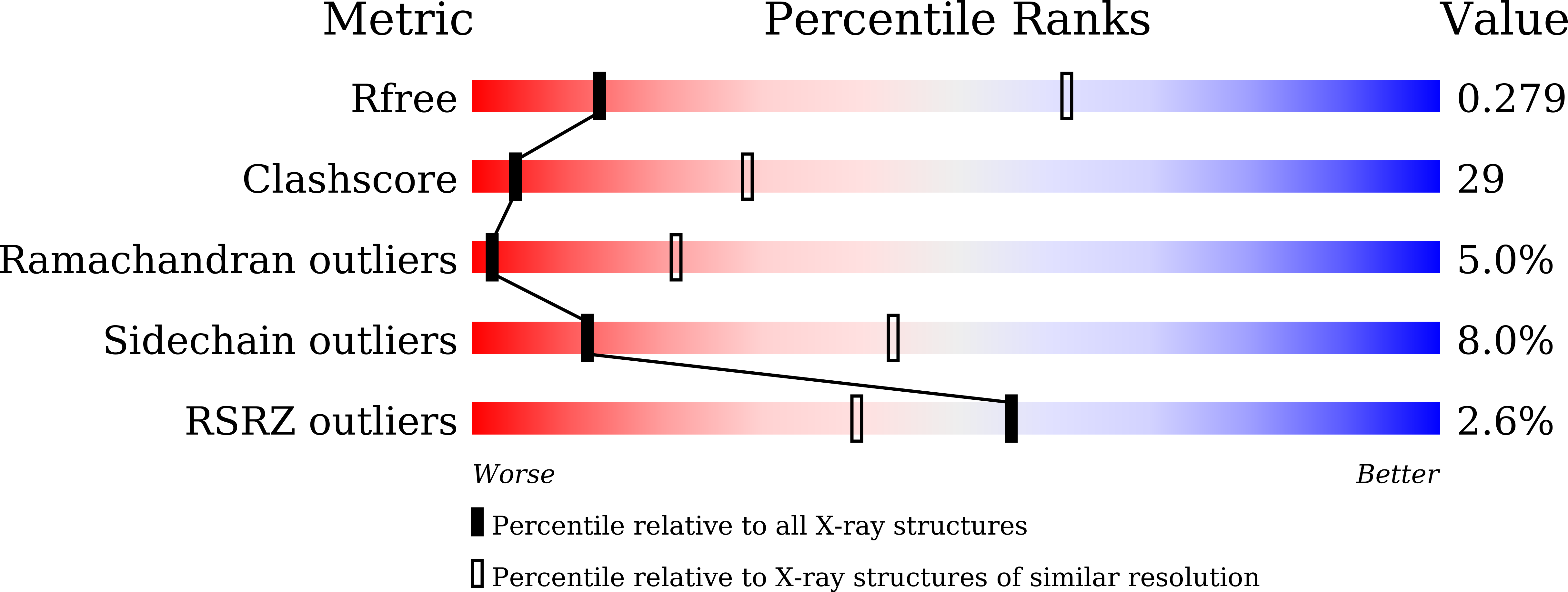
  • R-Value Free: 0.271 
  • R-Value Work: 0.207 
  • R-Value Observed: 0.209 

Starting Model: experimental
View more details

wwPDB Validation   3D Report Full Report


This is version 1.2 of the entry. See complete history


Literature

Beetle luciferases with naturally red- and blue-shifted emission.

Carrasco-Lopez, C.Ferreira, J.C.Lui, N.M.Schramm, S.Berraud-Pache, R.Navizet, I.Panjikar, S.Naumov, P.Rabeh, W.M.

(2018) Life Sci Alliance 1: e201800072-e201800072

  • DOI: https://doi.org/10.26508/lsa.201800072
  • Primary Citation of Related Structures:  
    6AAA, 6ABH, 6AC3

  • PubMed Abstract: 

    The different colors of light emitted by bioluminescent beetles that use an identical substrate and chemiexcitation reaction sequence to generate light remain a challenging and controversial mechanistic conundrum. The crystal structures of two beetle luciferases with red- and blue-shifted light relative to the green yellow light of the common firefly species provide direct insight into the molecular origin of the bioluminescence color. The structure of a blue-shifted green-emitting luciferase from the firefly Amydetes vivianii is monomeric with a structural fold similar to the previously reported firefly luciferases. The only known naturally red-emitting luciferase from the glow-worm Phrixothrix hirtus exists as tetramers and octamers. Structural and computational analyses reveal varying aperture between the two domains enclosing the active site. Mutagenesis analysis identified two conserved loops that contribute to the color of the emitted light. These results are expected to advance comparative computational studies into the conformational landscape of the luciferase reaction sequence.


  • Organizational Affiliation

    New York University Abu Dhabi, Abu Dhabi, United Arab Emirates.


Macromolecules
Find similar proteins by:  (by identity cutoff)  |  3D Structure
Entity ID: 1
MoleculeChains Sequence LengthOrganismDetailsImage
Red-bioluminescence eliciting luciferase
A, B, C, D
546Phrixothrix hirtusMutation(s): 0 
EC: 1.13.12.7
UniProt
Find proteins for Q9U4U7 (Phrixothrix hirtus)
Explore Q9U4U7 
Go to UniProtKB:  Q9U4U7
Entity Groups  
Sequence Clusters30% Identity50% Identity70% Identity90% Identity95% Identity100% Identity
UniProt GroupQ9U4U7
Sequence Annotations
Expand
  • Reference Sequence
Experimental Data & Validation

Experimental Data

  • Method: X-RAY DIFFRACTION
  • Resolution: 3.60 Å
  • R-Value Free: 0.271 
  • R-Value Work: 0.207 
  • R-Value Observed: 0.209 
  • Space Group: P 31 2 1
Unit Cell:
Length ( Å )Angle ( ˚ )
a = 119.1α = 90
b = 119.1β = 90
c = 351.402γ = 120
Software Package:
Software NamePurpose
PHENIXrefinement
iMOSFLMdata reduction
SCALAdata scaling
Auto-Rickshawphasing

Structure Validation

View Full Validation Report


Entry History & Funding Information

Deposition Data


Funding OrganizationLocationGrant Number
United Arab Emirates--

Revision History  (Full details and data files)

  • Version 1.0: 2018-08-22
    Type: Initial release
  • Version 1.1: 2018-12-05
    Changes: Data collection, Database references
  • Version 1.2: 2023-11-22
    Changes: Data collection, Database references, Refinement description