5XEQ

Crystal Structure of human MDGA1 and human neuroligin-2 complex


Experimental Data Snapshot

  • Method: X-RAY DIFFRACTION
  • Resolution: 3.14 Å
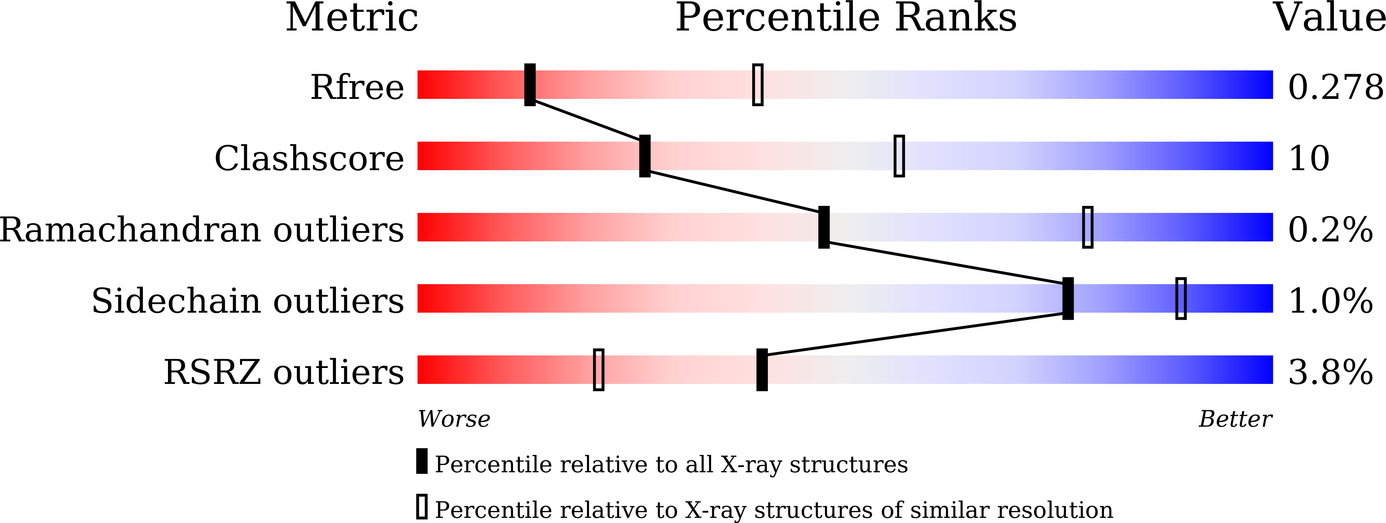
  • R-Value Free: 0.278 
  • R-Value Work: 0.234 
  • R-Value Observed: 0.238 

Starting Model: experimental
View more details

wwPDB Validation   3D Report Full Report


Ligand Structure Quality Assessment 


This is version 2.2 of the entry. See complete history


Literature

Structural Insights into Modulation of Neurexin-Neuroligin Trans-synaptic Adhesion by MDGA1/Neuroligin-2 Complex

Kim, J.A.Kim, D.Won, S.Y.Han, K.A.Park, D.Cho, E.Yun, N.An, H.J.Um, J.W.Kim, E.Lee, J.O.Ko, J.Kim, H.M.

(2017) Neuron 94: 1121-1131.e6

  • DOI: https://doi.org/10.1016/j.neuron.2017.05.034
  • Primary Citation of Related Structures:  
    5XEQ

  • PubMed Abstract: 

    Membrane-associated mucin domain-containing glycosylphosphatidylinositol anchor proteins (MDGAs) bind directly to neuroligin-1 (NL1) and neuroligin-2 (NL2), thereby respectively regulating excitatory and inhibitory synapse development. However, the mechanisms by which MDGAs modulate NL activity to specify development of the two synapse types remain unclear. Here, we determined the crystal structures of human NL2/MDGA1 Ig1-3 complex, revealing their stable 2:2 arrangement with three interaction interfaces. Cell-based assays using structure-guided, site-directed MDGA1 mutants showed that all three contact patches were required for the MDGA's negative regulation of NL2-mediated synaptogenic activity. Furthermore, MDGA1 competed with neurexins for NL2 via its Ig1 domain. The binding affinities of both MDGA1 and MDGA2 for NL1 and NL2 were similar, consistent with the structural prediction of similar binding interfaces. However, MDGA1 selectively associated with NL2, but not NL1, in vivo. These findings collectively provide structural insights into the mechanism by which MDGAs negatively modulate synapse development governed by NLs/neurexins.


  • Organizational Affiliation

    Graduate School of Nanoscience and Technology, Korea Advanced Institute of Science and Technology (KAIST), Daejeon 34141, Korea.


Macromolecules
Find similar proteins by:  (by identity cutoff)  |  3D Structure
Entity ID: 1
MoleculeChains Sequence LengthOrganismDetailsImage
Neuroligin-2576Homo sapiensMutation(s): 1 
Gene Names: NLGN2KIAA1366
UniProt & NIH Common Fund Data Resources
Find proteins for Q8NFZ4 (Homo sapiens)
Explore Q8NFZ4 
Go to UniProtKB:  Q8NFZ4
PHAROS:  Q8NFZ4
GTEx:  ENSG00000169992 
Entity Groups  
Sequence Clusters30% Identity50% Identity70% Identity90% Identity95% Identity100% Identity
UniProt GroupQ8NFZ4
Glycosylation
Glycosylation Sites: 1Go to GlyGen: Q8NFZ4-1
Sequence Annotations
Expand
  • Reference Sequence
Find similar proteins by:  (by identity cutoff)  |  3D Structure
Entity ID: 2
MoleculeChains Sequence LengthOrganismDetailsImage
MAM domain-containing glycosylphosphatidylinositol anchor protein 1319Homo sapiensMutation(s): 0 
Gene Names: MDGA1MAMDC3
UniProt & NIH Common Fund Data Resources
Find proteins for Q8NFP4 (Homo sapiens)
Explore Q8NFP4 
Go to UniProtKB:  Q8NFP4
PHAROS:  Q8NFP4
GTEx:  ENSG00000112139 
Entity Groups  
Sequence Clusters30% Identity50% Identity70% Identity90% Identity95% Identity100% Identity
UniProt GroupQ8NFP4
Glycosylation
Glycosylation Sites: 3Go to GlyGen: Q8NFP4-1
Sequence Annotations
Expand
  • Reference Sequence
Oligosaccharides

Help

Entity ID: 3
MoleculeChains Length2D Diagram Glycosylation3D Interactions
2-acetamido-2-deoxy-beta-D-glucopyranose-(1-4)-2-acetamido-2-deoxy-beta-D-glucopyranose
C
2N-Glycosylation
Glycosylation Resources
GlyTouCan:  G42666HT
GlyCosmos:  G42666HT
GlyGen:  G42666HT
Experimental Data & Validation

Experimental Data

  • Method: X-RAY DIFFRACTION
  • Resolution: 3.14 Å
  • R-Value Free: 0.278 
  • R-Value Work: 0.234 
  • R-Value Observed: 0.238 
  • Space Group: C 1 2 1
Unit Cell:
Length ( Å )Angle ( ˚ )
a = 154.875α = 90
b = 69.134β = 115.31
c = 108.843γ = 90
Software Package:
Software NamePurpose
PHENIXrefinement
SCALEPACKdata scaling
PDB_EXTRACTdata extraction
HKL-2000data reduction
PHASERphasing

Structure Validation

View Full Validation Report


Ligand Structure Quality Assessment 


Entry History 

Deposition Data

Revision History  (Full details and data files)

  • Version 1.0: 2017-07-12
    Type: Initial release
  • Version 1.1: 2017-08-30
    Changes: Data collection
  • Version 2.0: 2020-07-29
    Type: Remediation
    Reason: Carbohydrate remediation
    Changes: Atomic model, Data collection, Derived calculations, Structure summary
  • Version 2.1: 2023-11-22
    Changes: Data collection, Database references, Refinement description, Structure summary
  • Version 2.2: 2024-10-30
    Changes: Structure summary