5U8Q

Structure of the ectodomain of the human Type 1 insulin-like growth factor receptor in complex with IGF-I


Experimental Data Snapshot

  • Method: X-RAY DIFFRACTION
  • Resolution: 3.27 Å
  • R-Value Free: 0.303 
  • R-Value Work: 0.260 
  • R-Value Observed: 0.262 

Starting Model: experimental
View more details

wwPDB Validation   3D Report Full Report


Ligand Structure Quality Assessment 


This is version 2.1 of the entry. See complete history


Literature

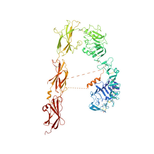
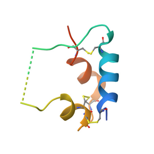
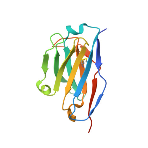
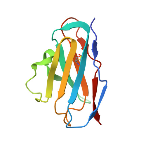
How ligand binds to the type 1 insulin-like growth factor receptor.

Xu, Y.Kong, G.K.Menting, J.G.Margetts, M.B.Delaine, C.A.Jenkin, L.M.Kiselyov, V.V.De Meyts, P.Forbes, B.E.Lawrence, M.C.

(2018) Nat Commun 9: 821-821

  • DOI: https://doi.org/10.1038/s41467-018-03219-7
  • Primary Citation of Related Structures:  
    5U8Q, 5U8R

  • PubMed Abstract: 

    Human type 1 insulin-like growth factor receptor is a homodimeric receptor tyrosine kinase that signals into pathways directing normal cellular growth, differentiation and proliferation, with aberrant signalling implicated in cancer. Insulin-like growth factor binding is understood to relax conformational restraints within the homodimer, initiating transphosphorylation of the tyrosine kinase domains. However, no three-dimensional structures exist for the receptor ectodomain to inform atomic-level understanding of these events. Here, we present crystal structures of the ectodomain in apo form and in complex with insulin-like growth factor I, the latter obtained by crystal soaking. These structures not only provide a wealth of detail of the growth factor interaction with the receptor's primary ligand-binding site but also indicate that ligand binding separates receptor domains by a mechanism of induced fit. Our findings are of importance to the design of agents targeting IGF-1R and its partner protein, the human insulin receptor.


  • Organizational Affiliation

    The Walter and Eliza Hall Institute of Medical Research, 1G Royal Parade, Parkville, VIC, 3052, Australia.


Macromolecules
Find similar proteins by:  (by identity cutoff)  |  3D Structure
Entity ID: 1
MoleculeChains Sequence LengthOrganismDetailsImage
Insulin-like growth factor 1 receptor885Homo sapiensMutation(s): 0 
Gene Names: IGF1R
EC: 2.7.10.1
UniProt & NIH Common Fund Data Resources
Find proteins for P08069 (Homo sapiens)
Explore P08069 
Go to UniProtKB:  P08069
GTEx:  ENSG00000140443 
Entity Groups  
Sequence Clusters30% Identity50% Identity70% Identity90% Identity95% Identity100% Identity
UniProt GroupP08069
Glycosylation
Glycosylation Sites: 6Go to GlyGen: P08069-1
Sequence Annotations
Expand
  • Reference Sequence
Find similar proteins by:  (by identity cutoff)  |  3D Structure
Entity ID: 2
MoleculeChains Sequence LengthOrganismDetailsImage
Insulin-like growth factor I70Homo sapiensMutation(s): 0 
Gene Names: IGF1IBP1
UniProt & NIH Common Fund Data Resources
Find proteins for P05019 (Homo sapiens)
Explore P05019 
Go to UniProtKB:  P05019
GTEx:  ENSG00000017427 
Entity Groups  
Sequence Clusters30% Identity50% Identity70% Identity90% Identity95% Identity100% Identity
UniProt GroupP05019
Sequence Annotations
Expand
  • Reference Sequence
Find similar proteins by:  (by identity cutoff)  |  3D Structure
Entity ID: 3
MoleculeChains Sequence LengthOrganismDetailsImage
Fv 24-60 heavy chainC [auth H]126Mus musculusMutation(s): 0 
Entity Groups  
Sequence Clusters30% Identity50% Identity70% Identity90% Identity95% Identity100% Identity
Sequence Annotations
Expand
  • Reference Sequence
Find similar proteins by:  (by identity cutoff)  |  3D Structure
Entity ID: 4
MoleculeChains Sequence LengthOrganismDetailsImage
Fv 24-60 light chainD [auth L]108Mus musculusMutation(s): 0 
Entity Groups  
Sequence Clusters30% Identity50% Identity70% Identity90% Identity95% Identity100% Identity
Sequence Annotations
Expand
  • Reference Sequence
Oligosaccharides

Help

Entity ID: 5
MoleculeChains Length2D Diagram Glycosylation3D Interactions
2-acetamido-2-deoxy-beta-D-glucopyranose-(1-4)-2-acetamido-2-deoxy-beta-D-glucopyranoseE [auth C],
G [auth E]
2N-Glycosylation
Glycosylation Resources
GlyTouCan:  G42666HT
GlyCosmos:  G42666HT
GlyGen:  G42666HT
Entity ID: 6
MoleculeChains Length2D Diagram Glycosylation3D Interactions
alpha-D-mannopyranose-(1-4)-alpha-D-mannopyranose-(1-4)-2-acetamido-2-deoxy-beta-D-glucopyranose-(1-4)-2-acetamido-2-deoxy-beta-D-glucopyranoseF [auth D]4N-Glycosylation
Glycosylation Resources
GlyTouCan:  G81874LD
GlyCosmos:  G81874LD
GlyGen:  G81874LD
Experimental Data & Validation

Experimental Data

  • Method: X-RAY DIFFRACTION
  • Resolution: 3.27 Å
  • R-Value Free: 0.303 
  • R-Value Work: 0.260 
  • R-Value Observed: 0.262 
  • Space Group: P 21 21 2
Unit Cell:
Length ( Å )Angle ( ˚ )
a = 88.69α = 90
b = 197.66β = 90
c = 117.65γ = 90
Software Package:
Software NamePurpose
phenix.refinerefinement
PHENIXrefinement
XDSdata reduction
XSCALEdata scaling
PHASERphasing

Structure Validation

View Full Validation Report


Ligand Structure Quality Assessment 


Entry History 

Deposition Data

Revision History  (Full details and data files)

  • Version 1.0: 2018-02-28
    Type: Initial release
  • Version 1.1: 2018-03-14
    Changes: Database references
  • Version 2.0: 2020-07-29
    Type: Remediation
    Reason: Carbohydrate remediation
    Changes: Atomic model, Data collection, Derived calculations, Structure summary
  • Version 2.1: 2023-10-04
    Changes: Data collection, Database references, Derived calculations, Refinement description, Structure summary