5OPY

Crystal structure of anti-alphaVbeta3 integrin Fab LM609


Experimental Data Snapshot

  • Method: X-RAY DIFFRACTION
  • Resolution: 2.26 Å
  • R-Value Free: 0.255 
  • R-Value Work: 0.213 
  • R-Value Observed: 0.215 

Starting Model: experimental
View more details

wwPDB Validation   3D Report Full Report


This is version 1.3 of the entry. See complete history


Literature

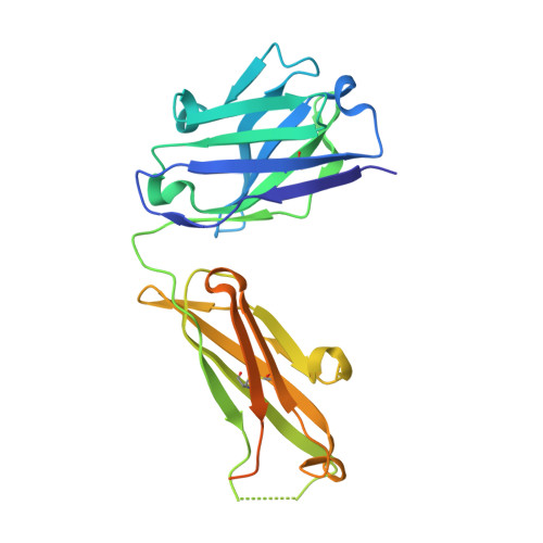
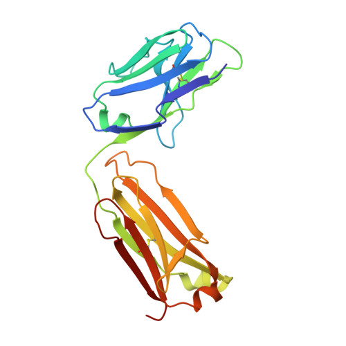
The Therapeutic Antibody LM609 Selectively Inhibits Ligand Binding to Human alpha V beta 3 Integrin via Steric Hindrance.

Borst, A.J.James, Z.M.Zagotta, W.N.Ginsberg, M.Rey, F.A.DiMaio, F.Backovic, M.Veesler, D.

(2017) Structure 25: 1732-1739.e5

  • DOI: https://doi.org/10.1016/j.str.2017.09.007
  • Primary Citation of Related Structures:  
    5OPY, 6AVQ, 6AVR, 6AVU

  • PubMed Abstract: 

    The LM609 antibody specifically recognizes α V β 3 integrin and inhibits angiogenesis, bone resorption, and viral infections in an arginine-glycine-aspartate-independent manner. LM609 entered phase II clinical trials for the treatment of several cancers and was also used for α V β 3 -targeted radioimmunotherapy. To elucidate the mechanisms of recognition and inhibition of α V β 3 integrin, we solved the structure of the LM609 antigen-binding fragment by X-ray crystallography and determined its binding affinity for α V β 3 . Using single-particle electron microscopy, we show that LM609 binds at the interface between the β-propeller domain of the α V chain and the βI domain of the β 3 chain, near the RGD-binding site, of all observed integrin conformational states. Integrating these data with fluorescence size-exclusion chromatography, we demonstrate that LM609 sterically hinders access of large ligands to the RGD-binding pocket, without obstructing it. This work provides a structural framework to expedite future efforts utilizing LM609 as a diagnostic or therapeutic tool.


  • Organizational Affiliation

    Department of Biochemistry, University of Washington, Seattle, WA 98195, USA.


Macromolecules
Find similar proteins by:  (by identity cutoff)  |  3D Structure
Entity ID: 1
MoleculeChains Sequence LengthOrganismDetailsImage
Heavy chain of LM609 Fab (antigen-binding fragment)A [auth H]257Mus musculusMutation(s): 0 
Entity Groups  
Sequence Clusters30% Identity50% Identity70% Identity90% Identity95% Identity100% Identity
Sequence Annotations
Expand
  • Reference Sequence
Find similar proteins by:  (by identity cutoff)  |  3D Structure
Entity ID: 2
MoleculeChains Sequence LengthOrganismDetailsImage
Light chain of LM609 Fab (antigen-binding fragment)B [auth L]214Mus musculusMutation(s): 0 
Entity Groups  
Sequence Clusters30% Identity50% Identity70% Identity90% Identity95% Identity100% Identity
Sequence Annotations
Expand
  • Reference Sequence
Experimental Data & Validation

Experimental Data

  • Method: X-RAY DIFFRACTION
  • Resolution: 2.26 Å
  • R-Value Free: 0.255 
  • R-Value Work: 0.213 
  • R-Value Observed: 0.215 
  • Space Group: P 2 21 21
Unit Cell:
Length ( Å )Angle ( ˚ )
a = 51.81α = 90
b = 82.35β = 90
c = 92.17γ = 90
Software Package:
Software NamePurpose
BUSTERrefinement
XDSdata reduction
Aimlessdata scaling
PHASERphasing

Structure Validation

View Full Validation Report


Entry History & Funding Information

Deposition Data


Funding OrganizationLocationGrant Number
Institut PasteurFranceRecurrent funding
National Institutes of Health/National Institute of General Medical Sciences (NIH/NIGMS)United States1R01GM120553
National Institutes of Health/National Institute of General Medical Sciences (NIH/NIGMS)United StatesT32GM008268

Revision History  (Full details and data files)

  • Version 1.0: 2017-10-25
    Type: Initial release
  • Version 1.1: 2017-11-15
    Changes: Database references
  • Version 1.2: 2022-03-30
    Changes: Author supporting evidence, Database references
  • Version 1.3: 2024-01-17
    Changes: Data collection, Refinement description