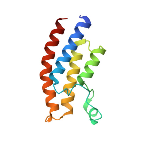
5O4T

Crystal structure of the human BRPF1 bromodomain in complex with BZ061


Experimental Data Snapshot

  • Method: X-RAY DIFFRACTION
  • Resolution: 1.50 Å
  • R-Value Free: 0.212 
  • R-Value Work: 0.182 
  • R-Value Observed: 0.185 

Starting Model: experimental
View more details

wwPDB Validation   3D Report Full Report


Ligand Structure Quality Assessment 


This is version 1.2 of the entry. See complete history


Literature

Structure-based discovery of selective BRPF1 bromodomain inhibitors.

Zhu, J.Zhou, C.Caflisch, A.

(2018) Eur J Med Chem 155: 337-352

  • DOI: https://doi.org/10.1016/j.ejmech.2018.05.037
  • Primary Citation of Related Structures:  
    5MWG, 5MWH, 5MWZ, 5O4S, 5O4T, 5O55, 5O5A, 5O5F, 5O5H, 5OV8, 5OWA, 6EKQ

  • PubMed Abstract: 

    Bromodomain and plant homeodomain (PHD) finger containing protein 1 (BRPF1) is a member of subfamily IV of the human bromodomains. Experimental evidence suggests that BRPF1 is involved in leukemia. In a previous high-throughput docking campaign we identified several chemotypes targeting the BRPF1 bromodomain. Here, pharmacophore searches using the binding modes of two of these chemotypes resulted in two new series of ligands of the BRPF1 bromodomain. The 2,3-dioxo-quinoxaline 21 exhibits a 2-μM affinity for the BRPF1 bromodomain in two different competition binding assays, and more than 100-fold selectivity for BRPF1 against other members of subfamily IV and representatives of other subfamilies. Cellular activity is confirmed by a viability assay in a leukemia cell line. Isothermal titration calorimetry measurements reveal enthalpy-driven binding for compounds 21, 26 (K D  = 3 μM), and the 2,4-dimethyl-oxazole derivative 42 (K D  = 10 μM). Multiple molecular dynamics simulations and a dozen co-crystal structures at high resolution provide useful information for further optimization of affinity for the BRPF1 bromodomain.


  • Organizational Affiliation

    Department of Biochemistry, University of Zurich, Winterthurerstrasse 190, CH-8057, Zurich, Switzerland.


Macromolecules
Find similar proteins by:  (by identity cutoff)  |  3D Structure
Entity ID: 1
MoleculeChains Sequence LengthOrganismDetailsImage
Peregrin116Homo sapiensMutation(s): 0 
Gene Names: BRPF1BR140
UniProt & NIH Common Fund Data Resources
Find proteins for P55201 (Homo sapiens)
Explore P55201 
Go to UniProtKB:  P55201
PHAROS:  P55201
GTEx:  ENSG00000156983 
Entity Groups  
Sequence Clusters30% Identity50% Identity70% Identity90% Identity95% Identity100% Identity
UniProt GroupP55201
Sequence Annotations
Expand
  • Reference Sequence
Small Molecules
Ligands 1 Unique
IDChains Name / Formula / InChI Key2D Diagram3D Interactions
9KT
Query on 9KT

Download Ideal Coordinates CCD File 
B [auth A]1,4-dimethylquinoxaline-2,3-dione
C10 H10 N2 O2
MXWJILNOXSNALO-UHFFFAOYSA-N
Binding Affinity Annotations 
IDSourceBinding Affinity
9KT BindingDB:  5O4T IC50: 4.00e+5 (nM) from 1 assay(s)
Experimental Data & Validation

Experimental Data

  • Method: X-RAY DIFFRACTION
  • Resolution: 1.50 Å
  • R-Value Free: 0.212 
  • R-Value Work: 0.182 
  • R-Value Observed: 0.185 
  • Space Group: P 32 2 1
Unit Cell:
Length ( Å )Angle ( ˚ )
a = 60.639α = 90
b = 60.639β = 90
c = 63.465γ = 120
Software Package:
Software NamePurpose
PHENIXrefinement
XDSdata reduction
Aimlessdata scaling
MOLREPphasing

Structure Validation

View Full Validation Report


Ligand Structure Quality Assessment 


Entry History 

Deposition Data

Revision History  (Full details and data files)

  • Version 1.0: 2018-06-13
    Type: Initial release
  • Version 1.1: 2018-06-27
    Changes: Data collection, Database references
  • Version 1.2: 2024-01-17
    Changes: Data collection, Database references, Refinement description