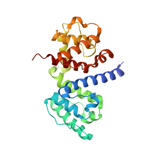
5NL7

The crystal structure of the Actin Binding Domain (ABD) of alpha actinin from Entamoeba histolytica


Experimental Data Snapshot

  • Method: X-RAY DIFFRACTION
  • Resolution: 2.48 Å
  • R-Value Free: 0.252 
  • R-Value Work: 0.196 
  • R-Value Observed: 0.199 

Starting Model: experimental
View more details

wwPDB Validation   3D Report Full Report


This is version 1.2 of the entry. See complete history


Literature

Calcium modulates the domain flexibility and function of an alpha-actinin similar to the ancestral alpha-actinin.

Pinotsis, N.Zielinska, K.Babuta, M.Arolas, J.L.Kostan, J.Khan, M.B.Schreiner, C.Salmazo, A.Ciccarelli, L.Puchinger, M.Gkougkoulia, E.A.Ribeiro Jr., E.A.Marlovits, T.C.Bhattacharya, A.Djinovic-Carugo, K.

(2020) Proc Natl Acad Sci U S A 117: 22101-22112

  • DOI: https://doi.org/10.1073/pnas.1917269117
  • Primary Citation of Related Structures:  
    5NL6, 5NL7, 6SL2, 6SL3, 6SL7

  • PubMed Abstract: 

    The actin cytoskeleton, a dynamic network of actin filaments and associated F-actin-binding proteins, is fundamentally important in eukaryotes. α-Actinins are major F-actin bundlers that are inhibited by Ca 2+ in nonmuscle cells. Here we report the mechanism of Ca 2+ -mediated regulation of Entamoeba histolytica α-actinin-2 ( Eh Actn2) with features expected for the common ancestor of Entamoeba and higher eukaryotic α-actinins. Crystal structures of Ca 2+ -free and Ca 2+ -bound Eh Actn2 reveal a calmodulin-like domain (CaMD) uniquely inserted within the rod domain. Integrative studies reveal an exceptionally high affinity of the Eh Actn2 CaMD for Ca 2+ , binding of which can only be regulated in the presence of physiological concentrations of Mg 2+ Ca 2+ binding triggers an increase in protein multidomain rigidity, reducing conformational flexibility of F-actin-binding domains via interdomain cross-talk and consequently inhibiting F-actin bundling. In vivo studies uncover that Eh Actn2 plays an important role in phagocytic cup formation and might constitute a new drug target for amoebic dysentery.


  • Organizational Affiliation

    Department of Structural and Computational Biology, Max Perutz Labs, University of Vienna, A-1030 Vienna, Austria.


Macromolecules
Find similar proteins by:  (by identity cutoff)  |  3D Structure
Entity ID: 1
MoleculeChains Sequence LengthOrganismDetailsImage
Calponin homology domain protein putative
A, B
234Entamoeba histolyticaMutation(s): 0 
Gene Names: CL6EHI_199000EHI_199000
UniProt
Find proteins for C4LWU6 (Entamoeba histolytica (strain ATCC 30459 / HM-1:IMSS / ABRM))
Explore C4LWU6 
Go to UniProtKB:  C4LWU6
Entity Groups  
Sequence Clusters30% Identity50% Identity70% Identity90% Identity95% Identity100% Identity
UniProt GroupC4LWU6
Sequence Annotations
Expand
  • Reference Sequence
Small Molecules
Ligands 2 Unique
IDChains Name / Formula / InChI Key2D Diagram3D Interactions
TRS
Query on TRS

Download Ideal Coordinates CCD File 
D [auth A]2-AMINO-2-HYDROXYMETHYL-PROPANE-1,3-DIOL
C4 H12 N O3
LENZDBCJOHFCAS-UHFFFAOYSA-O
CA
Query on CA

Download Ideal Coordinates CCD File 
C [auth A]CALCIUM ION
Ca
BHPQYMZQTOCNFJ-UHFFFAOYSA-N
Experimental Data & Validation

Experimental Data

  • Method: X-RAY DIFFRACTION
  • Resolution: 2.48 Å
  • R-Value Free: 0.252 
  • R-Value Work: 0.196 
  • R-Value Observed: 0.199 
  • Space Group: P 3
Unit Cell:
Length ( Å )Angle ( ˚ )
a = 146.74α = 90
b = 146.74β = 90
c = 33.97γ = 120
Software Package:
Software NamePurpose
PHENIXrefinement
XDSdata reduction
XSCALEdata scaling
MOLREPphasing

Structure Validation

View Full Validation Report


Entry History 

Deposition Data

Revision History  (Full details and data files)

  • Version 1.0: 2018-05-16
    Type: Initial release
  • Version 1.1: 2020-11-04
    Changes: Database references, Derived calculations
  • Version 1.2: 2024-01-17
    Changes: Data collection, Database references, Refinement description