5MWL

INOSITOL 1,3,4,5,6-PENTAKISPHOSPHATE 2-KINASE FROM M. MUSCULUS IN COMPLEX WITH ATP and IP5


Experimental Data Snapshot

  • Method: X-RAY DIFFRACTION
  • Resolution: 3.20 Å
  • R-Value Free: 0.245 
  • R-Value Work: 0.225 
  • R-Value Observed: 0.226 

Starting Model: experimental
View more details

wwPDB Validation   3D Report Full Report


Ligand Structure Quality Assessment 


This is version 1.3 of the entry. See complete history


Literature

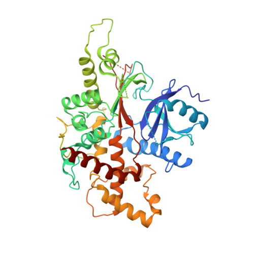
The crystal structure of mammalian inositol 1,3,4,5,6-pentakisphosphate 2-kinase reveals a new zinc-binding site and key features for protein function.

Franco-Echevarria, E.Sanz-Aparicio, J.Brearley, C.A.Gonzalez-Rubio, J.M.Gonzalez, B.

(2017) J Biol Chem 292: 10534-10548

  • DOI: https://doi.org/10.1074/jbc.M117.780395
  • Primary Citation of Related Structures:  
    5MW8, 5MWL, 5MWM

  • PubMed Abstract: 

    Inositol 1,3,4,5,6-pentakisphosphate 2-kinases (IP 5 2-Ks) are part of a family of enzymes in charge of synthesizing inositol hexakisphosphate (IP 6 ) in eukaryotic cells. This protein and its product IP 6 present many roles in cells, participating in mRNA export, embryonic development, and apoptosis. We reported previously that the full-length IP 5 2-K from Arabidopsis thaliana is a zinc metallo-enzyme, including two separated lobes (the N- and C-lobes). We have also shown conformational changes in IP 5 2-K and have identified the residues involved in substrate recognition and catalysis. However, the specific features of mammalian IP 5 2-Ks remain unknown. To this end, we report here the first structure for a murine IP 5 2-K in complex with ATP/IP 5 or IP 6 Our structural findings indicated that the general folding in N- and C-lobes is conserved with A. thaliana IP 5 2-K. A helical scaffold in the C-lobe constitutes the inositol phosphate-binding site, which, along with the participation of the N-lobe, endows high specificity to this protein. However, we also noted large structural differences between the orthologues from these two eukaryotic kingdoms. These differences include a novel zinc-binding site and regions unique to the mammalian IP 5 2-K, as an unexpected basic patch on the protein surface. In conclusion, our findings have uncovered distinct features of a mammalian IP 5 2-K and set the stage for investigations into protein-protein or protein-RNA interactions important for IP 5 2-K function and activity.


  • Organizational Affiliation

    From the Departamento de Cristalografía y Biología Estructural, Instituto de Química-Física "Rocasolano," Consejo Superior de Investigaciones Científicas, Serrano 119, 28006 Madrid, Spain and.


Macromolecules
Find similar proteins by:  (by identity cutoff)  |  3D Structure
Entity ID: 1
MoleculeChains Sequence LengthOrganismDetailsImage
Inositol-pentakisphosphate 2-kinase
A, B
471Mus musculusMutation(s): 0 
Gene Names: Ippk
EC: 2.7.1.158
UniProt & NIH Common Fund Data Resources
Find proteins for Q6P1C1 (Mus musculus)
Explore Q6P1C1 
Go to UniProtKB:  Q6P1C1
IMPC:  MGI:1922928
Entity Groups  
Sequence Clusters30% Identity50% Identity70% Identity90% Identity95% Identity100% Identity
UniProt GroupQ6P1C1
Sequence Annotations
Expand
  • Reference Sequence
Small Molecules
Ligands 4 Unique
IDChains Name / Formula / InChI Key2D Diagram3D Interactions
5MY
Query on 5MY

Download Ideal Coordinates CCD File 
D [auth A]MYO-INOSITOL-(1,3,4,5,6)-PENTAKISPHOSPHATE
C6 H17 O21 P5
CTPQAXVNYGZUAJ-KXXVROSKSA-N
ATP
Query on ATP

Download Ideal Coordinates CCD File 
C [auth A]ADENOSINE-5'-TRIPHOSPHATE
C10 H16 N5 O13 P3
ZKHQWZAMYRWXGA-KQYNXXCUSA-N
ZN
Query on ZN

Download Ideal Coordinates CCD File 
E [auth A],
F [auth A],
I [auth B],
J [auth B]
ZINC ION
Zn
PTFCDOFLOPIGGS-UHFFFAOYSA-N
MG
Query on MG

Download Ideal Coordinates CCD File 
G [auth A],
H [auth A]
MAGNESIUM ION
Mg
JLVVSXFLKOJNIY-UHFFFAOYSA-N
Experimental Data & Validation

Experimental Data

  • Method: X-RAY DIFFRACTION
  • Resolution: 3.20 Å
  • R-Value Free: 0.245 
  • R-Value Work: 0.225 
  • R-Value Observed: 0.226 
  • Space Group: P 1 21 1
Unit Cell:
Length ( Å )Angle ( ˚ )
a = 64.623α = 90
b = 140.758β = 106.53
c = 68.662γ = 90
Software Package:
Software NamePurpose
REFMACrefinement
XDSdata reduction
XDSdata scaling
Aimlessdata scaling
Cootmodel building

Structure Validation

View Full Validation Report


Ligand Structure Quality Assessment 


Entry History & Funding Information

Deposition Data


Funding OrganizationLocationGrant Number
CSICSpainBFU2014-53762-P

Revision History  (Full details and data files)

  • Version 1.0: 2017-05-10
    Type: Initial release
  • Version 1.1: 2017-07-05
    Changes: Database references
  • Version 1.2: 2019-10-16
    Changes: Data collection
  • Version 1.3: 2024-01-17
    Changes: Data collection, Database references, Derived calculations, Refinement description