5MIO

KIF2C-DARPIN FUSION PROTEIN BOUND TO TUBULIN


Experimental Data Snapshot

  • Method: X-RAY DIFFRACTION
  • Resolution: 3.19 Å
  • R-Value Free: 0.257 
  • R-Value Work: 0.211 
  • R-Value Observed: 0.213 

Starting Models: experimental
View more details

wwPDB Validation   3D Report Full Report


Ligand Structure Quality Assessment 


This is version 1.3 of the entry. See complete history


Literature

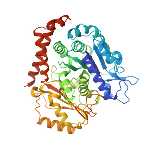
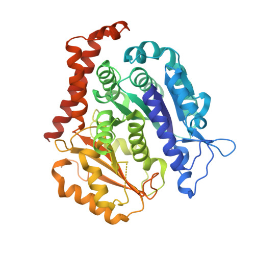
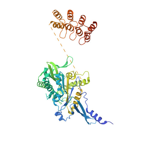
Insight into microtubule disassembly by kinesin-13s from the structure of Kif2C bound to tubulin.

Wang, W.Cantos-Fernandes, S.Lv, Y.Kuerban, H.Ahmad, S.Wang, C.Gigant, B.

(2017) Nat Commun 8: 70-70

  • DOI: https://doi.org/10.1038/s41467-017-00091-9
  • Primary Citation of Related Structures:  
    5MIO

  • PubMed Abstract: 

    Kinesin-13s are critical microtubule regulators which induce microtubule disassembly in an ATP dependent manner. To clarify their mechanism, we report here the crystal structure of a functional construct of the kinesin-13 Kif2C/MCAK in an ATP-like state and bound to the αβ-tubulin heterodimer, a complex mimicking the species that dissociates from microtubule ends during catalytic disassembly. Our results picture how Kif2C stabilizes a curved tubulin conformation. The Kif2C α4-L12-α5 region undergoes a remarkable 25° rotation upon tubulin binding to target the αβ-tubulin hinge. This movement leads the β5a-β5b motif to interact with the distal end of β-tubulin, whereas the neck and the KVD motif, two specific elements of kinesin-13s, target the α-tubulin distal end. Taken together with the study of Kif2C mutants, our data suggest that stabilization of a curved tubulin is an important contribution to the Kif2C mechanism.Kinesin-13s are microtubule depolymerizing enzymes. Here the authors present the crystal structure of a DARPin fused construct comprising the short neck region and motor domain of kinesin-13 in complex with an αβ-tubulin heterodimer, which shows that kinesin-13 functions by stabilizing a curved tubulin conformation.


  • Organizational Affiliation

    Department of Central Laboratory, Shanghai Tenth People's Hospital of Tongji University, School of Life Sciences and Technology, Tongji University, Shanghai, 200092, China.


Macromolecules
Find similar proteins by:  (by identity cutoff)  |  3D Structure
Entity ID: 1
MoleculeChains Sequence LengthOrganismDetailsImage
Tubulin alpha-1B chain451Bos taurusMutation(s): 0 
UniProt
Find proteins for P81947 (Bos taurus)
Explore P81947 
Go to UniProtKB:  P81947
Entity Groups  
Sequence Clusters30% Identity50% Identity70% Identity90% Identity95% Identity100% Identity
UniProt GroupP81947
Sequence Annotations
Expand
  • Reference Sequence
Find similar proteins by:  (by identity cutoff)  |  3D Structure
Entity ID: 2
MoleculeChains Sequence LengthOrganismDetailsImage
Tubulin beta chain445Ovis ariesMutation(s): 0 
UniProt
Find proteins for D0VWY9 (Ovis aries)
Explore D0VWY9 
Go to UniProtKB:  D0VWY9
Entity Groups  
Sequence Clusters30% Identity50% Identity70% Identity90% Identity95% Identity100% Identity
UniProt GroupD0VWY9
Sequence Annotations
Expand
  • Reference Sequence
Find similar proteins by:  (by identity cutoff)  |  3D Structure
Entity ID: 3
MoleculeChains Sequence LengthOrganismDetailsImage
Kinesin-like protein KIF2C,KIF2C FUSED TO A DARPIN,KIF2C FUSED TO A DARPIN573Homo sapiensMutation(s): 2 
Gene Names: KIF2CKNSL6
UniProt & NIH Common Fund Data Resources
Find proteins for Q99661 (Homo sapiens)
Explore Q99661 
Go to UniProtKB:  Q99661
GTEx:  ENSG00000142945 
Entity Groups  
Sequence Clusters30% Identity50% Identity70% Identity90% Identity95% Identity100% Identity
UniProt GroupQ99661
Sequence Annotations
Expand
  • Reference Sequence
Small Molecules
Ligands 5 Unique
IDChains Name / Formula / InChI Key2D Diagram3D Interactions
GTP
Query on GTP

Download Ideal Coordinates CCD File 
D [auth A]GUANOSINE-5'-TRIPHOSPHATE
C10 H16 N5 O14 P3
XKMLYUALXHKNFT-UUOKFMHZSA-N
ANP
Query on ANP

Download Ideal Coordinates CCD File 
I [auth C]PHOSPHOAMINOPHOSPHONIC ACID-ADENYLATE ESTER
C10 H17 N6 O12 P3
PVKSNHVPLWYQGJ-KQYNXXCUSA-N
GDP
Query on GDP

Download Ideal Coordinates CCD File 
F [auth B]GUANOSINE-5'-DIPHOSPHATE
C10 H15 N5 O11 P2
QGWNDRXFNXRZMB-UUOKFMHZSA-N
LOC
Query on LOC

Download Ideal Coordinates CCD File 
G [auth B]N-[(7S)-1,2,3,10-tetramethoxy-9-oxo-6,7-dihydro-5H-benzo[d]heptalen-7-yl]ethanamide
C22 H25 N O6
IAKHMKGGTNLKSZ-INIZCTEOSA-N
MG
Query on MG

Download Ideal Coordinates CCD File 
E [auth A],
H [auth C]
MAGNESIUM ION
Mg
JLVVSXFLKOJNIY-UHFFFAOYSA-N
Binding Affinity Annotations 
IDSourceBinding Affinity
LOC BindingDB:  5MIO Ki: min: 1.6, max: 5750 (nM) from 4 assay(s)
Kd: min: 3700, max: 1.13e+4 (nM) from 2 assay(s)
IC50: min: 6.7, max: 2.93e+4 (nM) from 24 assay(s)
Experimental Data & Validation

Experimental Data

  • Method: X-RAY DIFFRACTION
  • Resolution: 3.19 Å
  • R-Value Free: 0.257 
  • R-Value Work: 0.211 
  • R-Value Observed: 0.213 
  • Space Group: C 2 2 21
Unit Cell:
Length ( Å )Angle ( ˚ )
a = 51.81α = 90
b = 229.76β = 90
c = 293.95γ = 90
Software Package:
Software NamePurpose
BUSTERrefinement
XDSdata reduction
Aimlessdata scaling
PHASERphasing

Structure Validation

View Full Validation Report


Ligand Structure Quality Assessment 


Entry History & Funding Information

Deposition Data


Funding OrganizationLocationGrant Number
French National Research AgencyFranceANR-12-BSV8-0002-01
Fondation ARCFrancePDF 20130606987

Revision History  (Full details and data files)

  • Version 1.0: 2017-07-19
    Type: Initial release
  • Version 1.1: 2017-09-06
    Changes: Author supporting evidence
  • Version 1.2: 2019-06-12
    Changes: Data collection, Structure summary
  • Version 1.3: 2024-01-17
    Changes: Data collection, Database references, Derived calculations, Refinement description