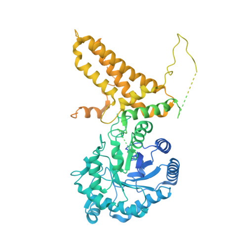
5M7S

Structure of human O-GlcNAc hydrolase with bound transition state analog ThiametG


Experimental Data Snapshot

  • Method: X-RAY DIFFRACTION
  • Resolution: 2.40 Å
  • R-Value Free: 0.232 
  • R-Value Work: 0.207 
  • R-Value Observed: 0.208 

wwPDB Validation   3D Report Full Report


Ligand Structure Quality Assessment 


This is version 1.3 of the entry. See complete history


Literature

Structural and functional insight into human O-GlcNAcase.

Roth, C.Chan, S.Offen, W.A.Hemsworth, G.R.Willems, L.I.King, D.T.Varghese, V.Britton, R.Vocadlo, D.J.Davies, G.J.

(2017) Nat Chem Biol 13: 610-612

  • DOI: https://doi.org/10.1038/nchembio.2358
  • Primary Citation of Related Structures:  
    5M7R, 5M7S, 5M7T, 5M7U

  • PubMed Abstract: 

    O-GlcNAc hydrolase (OGA) removes O-linked N-acetylglucosamine (O-GlcNAc) from a myriad of nucleocytoplasmic proteins. Through co-expression and assembly of OGA fragments, we determined the three-dimensional structure of human OGA, revealing an unusual helix-exchanged dimer that lays a structural foundation for an improved understanding of substrate recognition and regulation of OGA. Structures of OGA in complex with a series of inhibitors define a precise blueprint for the design of inhibitors that have clinical value.


  • Organizational Affiliation

    York Structural Biology Laboratory, Department of Chemistry University of York, York, UK.


Macromolecules
Find similar proteins by:  (by identity cutoff)  |  3D Structure
Entity ID: 1
MoleculeChains Sequence LengthOrganismDetailsImage
Protein O-GlcNAcase
A, B
916Homo sapiensMutation(s): 0 
Gene Names: MGEA5HEXCKIAA0679MEA5
EC: 3.2.1.169 (PDB Primary Data), 3.2.1 (PDB Primary Data)
UniProt & NIH Common Fund Data Resources
Find proteins for O60502 (Homo sapiens)
Explore O60502 
Go to UniProtKB:  O60502
PHAROS:  O60502
GTEx:  ENSG00000198408 
Entity Groups  
Sequence Clusters30% Identity50% Identity70% Identity90% Identity95% Identity100% Identity
UniProt GroupO60502
Sequence Annotations
Expand
  • Reference Sequence
Small Molecules
Ligands 1 Unique
IDChains Name / Formula / InChI Key2D Diagram3D Interactions
NHT
Query on NHT

Download Ideal Coordinates CCD File 
C [auth A],
D [auth B]
(3AR,5R,6S,7R,7AR)-2-(ETHYLAMINO)-5-(HYDROXYMETHYL)-5,6,7,7A-TETRAHYDRO-3AH-PYRANO[3,2-D][1,3]THIAZOLE-6,7-DIOL
C9 H16 N2 O4 S
PPAIMZHKIXDJRN-FMDGEEDCSA-N
Binding Affinity Annotations 
IDSourceBinding Affinity
NHT BindingDB:  5M7S Ki: min: 0.4, max: 21 (nM) from 4 assay(s)
IC50: min: 0.41, max: 3 (nM) from 2 assay(s)
EC50: min: 13, max: 30 (nM) from 4 assay(s)
Experimental Data & Validation

Experimental Data

  • Method: X-RAY DIFFRACTION
  • Resolution: 2.40 Å
  • R-Value Free: 0.232 
  • R-Value Work: 0.207 
  • R-Value Observed: 0.208 
  • Space Group: P 43 21 2
Unit Cell:
Length ( Å )Angle ( ˚ )
a = 101.908α = 90
b = 101.908β = 90
c = 282.799γ = 90
Software Package:
Software NamePurpose
REFMACrefinement
PDB_EXTRACTdata extraction
REFMACrefinement
XDSdata reduction
Aimlessdata scaling
REFMACphasing

Structure Validation

View Full Validation Report


Ligand Structure Quality Assessment 


Entry History & Funding Information

Deposition Data


Funding OrganizationLocationGrant Number
Biotechnology and Biological Sciences Research CouncilUnited KingdomBB/K003836/1
Canadian Institutes of Health ResearchCanadaMOP-123341

Revision History  (Full details and data files)

  • Version 1.0: 2017-03-29
    Type: Initial release
  • Version 1.1: 2017-04-05
    Changes: Database references
  • Version 1.2: 2017-05-24
    Changes: Database references
  • Version 1.3: 2017-08-30
    Changes: Author supporting evidence