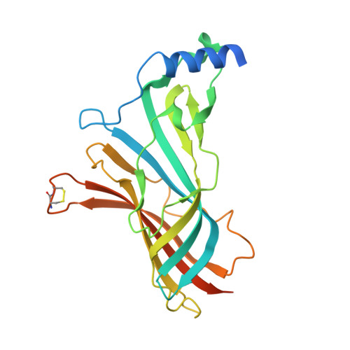
5LXB

Crystal structure of a mutant binding protein (5HTBP-AChBP) in complex with palonosetron


Experimental Data Snapshot

  • Method: X-RAY DIFFRACTION
  • Resolution: 2.34 Å
  • R-Value Free: 0.245 
  • R-Value Work: 0.218 
  • R-Value Observed: 0.219 

Starting Model: experimental
View more details

wwPDB Validation   3D Report Full Report


Ligand Structure Quality Assessment 


This is version 1.3 of the entry. See complete history


Literature

Palonosetron-5-HT3 Receptor Interactions As Shown by a Binding Protein Cocrystal Structure.

Price, K.L.Lillestol, R.K.Ulens, C.Lummis, S.C.

(2016) ACS Chem Neurosci 7: 1641-1646

  • DOI: https://doi.org/10.1021/acschemneuro.6b00132
  • Primary Citation of Related Structures:  
    5LXB

  • PubMed Abstract: 

    Palonosetron is a potent 5-HT 3 receptor antagonist and an effective therapeutic agent against emesis. Here we identify the molecular determinants of compound recognition in the receptor binding site by obtaining a high resolution structure of palonosetron bound to an engineered acetylcholine binding protein that mimics the 5-HT 3 receptor binding site, termed 5-HTBP, and by examining the potency of palonosetron in a range of 5-HT 3 receptors with mutated binding site residues. The structural data indicate that palonosetron forms a tight and effective wedge in the binding pocket, made possible by its rigid tricyclic ring structure and its interactions with binding site residues; it adopts a binding pose that is distinct from the related antiemetics granisetron and tropisetron. The functional data show many residues previously shown to interact with agonists and antagonists in the binding site are important for palonosetron binding, and indicate those of particular importance are W183 (a cation-π interaction and a hydrogen bond) and Y153 (a hydrogen bond). This information, and the availability of the structure of palonosetron bound to 5-HTBP, should aid the development of novel and more efficacious drugs that act via 5-HT 3 receptors.


  • Organizational Affiliation

    Department of Biochemistry, University of Cambridge , Tennis Court Road, Cambridge CB2 1QW, United Kingdom.


Macromolecules
Find similar proteins by:  (by identity cutoff)  |  3D Structure
Entity ID: 1
MoleculeChains Sequence LengthOrganismDetailsImage
Soluble acetylcholine receptor
A, B, C, D, E
A, B, C, D, E, F, G, H, I, J
242Aplysia californicaMutation(s): 0 
UniProt
Find proteins for Q8WSF8 (Aplysia californica)
Explore Q8WSF8 
Go to UniProtKB:  Q8WSF8
Entity Groups  
Sequence Clusters30% Identity50% Identity70% Identity90% Identity95% Identity100% Identity
UniProt GroupQ8WSF8
Sequence Annotations
Expand
  • Reference Sequence
Small Molecules
Ligands 2 Unique
IDChains Name / Formula / InChI Key2D Diagram3D Interactions
7A9
Query on 7A9

Download Ideal Coordinates CCD File 
L [auth B]
N [auth C]
P [auth D]
R [auth E]
U [auth G]
L [auth B],
N [auth C],
P [auth D],
R [auth E],
U [auth G],
V [auth H],
Y [auth J]
palonosetron
C19 H24 N2 O
CPZBLNMUGSZIPR-DOTOQJQBSA-N
NAG
Query on NAG

Download Ideal Coordinates CCD File 
K [auth A]
M [auth C]
O [auth D]
Q [auth E]
S [auth F]
K [auth A],
M [auth C],
O [auth D],
Q [auth E],
S [auth F],
T [auth G],
W [auth I],
X [auth J]
2-acetamido-2-deoxy-beta-D-glucopyranose
C8 H15 N O6
OVRNDRQMDRJTHS-FMDGEEDCSA-N
Experimental Data & Validation

Experimental Data

  • Method: X-RAY DIFFRACTION
  • Resolution: 2.34 Å
  • R-Value Free: 0.245 
  • R-Value Work: 0.218 
  • R-Value Observed: 0.219 
  • Space Group: P 1 21 1
Unit Cell:
Length ( Å )Angle ( ˚ )
a = 72.215α = 90
b = 137.051β = 93.78
c = 131.249γ = 90
Software Package:
Software NamePurpose
REFMACrefinement
PDB_EXTRACTdata extraction
SCALAdata scaling
MOLREPphasing

Structure Validation

View Full Validation Report


Ligand Structure Quality Assessment 


Entry History & Funding Information

Deposition Data

  • Released Date: 2016-10-12 
  • Deposition Author(s): Ulens, C.

Funding OrganizationLocationGrant Number
KU LeuvenBelgiumOT/13/095

Revision History  (Full details and data files)

  • Version 1.0: 2016-10-12
    Type: Initial release
  • Version 1.1: 2017-01-11
    Changes: Database references
  • Version 1.2: 2020-07-29
    Type: Remediation
    Reason: Carbohydrate remediation
    Changes: Data collection, Derived calculations, Structure summary
  • Version 1.3: 2024-01-17
    Changes: Data collection, Database references, Refinement description, Structure summary