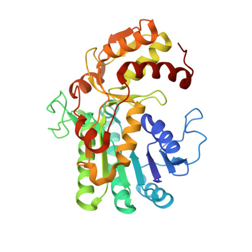
5LC1

L-threonine dehydrogenase from Trypanosoma brucei with NAD and the inhibitor pyruvate bound.


Experimental Data Snapshot

  • Method: X-RAY DIFFRACTION
  • Resolution: 2.10 Å
  • R-Value Free: 0.208 
  • R-Value Work: 0.147 
  • R-Value Observed: 0.150 

Starting Model: experimental
View more details

wwPDB Validation   3D Report Full Report


Ligand Structure Quality Assessment 


This is version 2.2 of the entry. See complete history


Literature

Structure and function of L-threonine-3-dehydrogenase from the parasitic protozoan Trypanosoma brucei revealed by X-ray crystallography and geometric simulations.

Adjogatse, E.Erskine, P.Wells, S.A.Kelly, J.M.Wilden, J.D.Chan, A.W.E.Selwood, D.Coker, A.Wood, S.Cooper, J.B.

(2018) Acta Crystallogr D Struct Biol 74: 861-876

  • DOI: https://doi.org/10.1107/S2059798318009208
  • Primary Citation of Related Structures:  
    5K4Q, 5K4T, 5K4U, 5K4V, 5K4W, 5K4Y, 5K50, 5L9A, 5LC1

  • PubMed Abstract: 

    Two of the world's most neglected tropical diseases, human African trypanosomiasis (HAT) and Chagas disease, are caused by protozoan parasites of the genus Trypanosoma. These organisms possess specialized metabolic pathways, frequently distinct from those in humans, which have potential to be exploited as novel drug targets. This study elucidates the structure and function of L-threonine-3-dehydrogenase (TDH) from T. brucei, the causative pathogen of HAT. TDH is a key enzyme in the metabolism of L-threonine, and an inhibitor of TDH has been shown to have trypanocidal activity in the procyclic form of T. brucei. TDH is a nonfunctional pseudogene in humans, suggesting that it may be possible to rationally design safe and specific therapies for trypanosomiasis by targeting this parasite enzyme. As an initial step, the TDH gene from T. brucei was expressed and the three-dimensional structure of the enzyme was solved by X-ray crystallography. In multiple crystallographic structures, T. brucei TDH is revealed to be a dimeric short-chain dehydrogenase that displays a considerable degree of conformational variation in its ligand-binding regions. Geometric simulations of the structure have provided insight into the dynamic behaviour of this enzyme. Furthermore, structures of TDH bound to its natural substrates and known inhibitors have been determined, giving an indication of the mechanism of catalysis of the enzyme. Collectively, these results provide vital details for future drug design to target TDH or related enzymes.


  • Organizational Affiliation

    Laboratory for Protein Crystallography, Division of Medicine, University College London, Gower Street, London WC1E 7HT, England.


Macromolecules
Find similar proteins by:  (by identity cutoff)  |  3D Structure
Entity ID: 1
MoleculeChains Sequence LengthOrganismDetailsImage
L-threonine 3-dehydrogenase
A, B, C, D, E
A, B, C, D, E, F
321Trypanosoma bruceiMutation(s): 0 
EC: 1.1.1.103
UniProt
Find proteins for Q7YW97 (Trypanosoma brucei)
Explore Q7YW97 
Go to UniProtKB:  Q7YW97
Entity Groups  
Sequence Clusters30% Identity50% Identity70% Identity90% Identity95% Identity100% Identity
UniProt GroupQ7YW97
Sequence Annotations
Expand
  • Reference Sequence
Small Molecules
Ligands 5 Unique
IDChains Name / Formula / InChI Key2D Diagram3D Interactions
NAD
Query on NAD

Download Ideal Coordinates CCD File 
AA [auth F]
G [auth A]
L [auth B]
Q [auth C]
T [auth D]
AA [auth F],
G [auth A],
L [auth B],
Q [auth C],
T [auth D],
W [auth E]
NICOTINAMIDE-ADENINE-DINUCLEOTIDE
C21 H27 N7 O14 P2
BAWFJGJZGIEFAR-NNYOXOHSSA-N
PYR
Query on PYR

Download Ideal Coordinates CCD File 
BA [auth F]
H [auth A]
M [auth B]
R [auth C]
U [auth D]
BA [auth F],
H [auth A],
M [auth B],
R [auth C],
U [auth D],
X [auth E]
PYRUVIC ACID
C3 H4 O3
LCTONWCANYUPML-UHFFFAOYSA-N
BME
Query on BME

Download Ideal Coordinates CCD File 
I [auth A],
N [auth B]
BETA-MERCAPTOETHANOL
C2 H6 O S
DGVVWUTYPXICAM-UHFFFAOYSA-N
ACT
Query on ACT

Download Ideal Coordinates CCD File 
J [auth A],
O [auth B],
Y [auth E]
ACETATE ION
C2 H3 O2
QTBSBXVTEAMEQO-UHFFFAOYSA-M
NA
Query on NA

Download Ideal Coordinates CCD File 
CA [auth F]
K [auth A]
P [auth B]
S [auth C]
V [auth D]
CA [auth F],
K [auth A],
P [auth B],
S [auth C],
V [auth D],
Z [auth E]
SODIUM ION
Na
FKNQFGJONOIPTF-UHFFFAOYSA-N
Experimental Data & Validation

Experimental Data

  • Method: X-RAY DIFFRACTION
  • Resolution: 2.10 Å
  • R-Value Free: 0.208 
  • R-Value Work: 0.147 
  • R-Value Observed: 0.150 
  • Space Group: P 21 21 2
Unit Cell:
Length ( Å )Angle ( ˚ )
a = 132.04α = 90
b = 276.49β = 90
c = 55.74γ = 90
Software Package:
Software NamePurpose
MOSFLMdata reduction
MOLREPphasing
REFMACrefinement

Structure Validation

View Full Validation Report


Ligand Structure Quality Assessment 


Entry History 

Deposition Data

Revision History  (Full details and data files)

  • Version 1.0: 2016-07-27
    Type: Initial release
  • Version 1.1: 2018-09-19
    Changes: Data collection, Database references
  • Version 1.2: 2018-10-10
    Changes: Data collection, Database references
  • Version 2.0: 2023-11-15
    Changes: Atomic model, Data collection, Database references, Derived calculations
  • Version 2.1: 2024-01-10
    Changes: Refinement description
  • Version 2.2: 2024-04-03
    Changes: Derived calculations