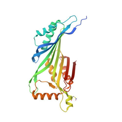
5K9G

Crystal Structure of GTP Cyclohydrolase-IB with Tris


Experimental Data Snapshot

  • Method: X-RAY DIFFRACTION
  • Resolution: 1.90 Å
  • R-Value Free: 0.221 
  • R-Value Work: 0.173 
  • R-Value Observed: 0.175 

Starting Model: experimental
View more details

wwPDB Validation   3D Report Full Report


This is version 1.5 of the entry. See complete history


Literature

Mechanism and catalytic strategy of the prokaryotic-specific GTP cyclohydrolase-IB.

Paranagama, N.Bonnett, S.A.Alvarez, J.Luthra, A.Stec, B.Gustafson, A.Iwata-Reuyl, D.Swairjo, M.A.

(2017) Biochem J 474: 1017-1039

  • DOI: https://doi.org/10.1042/BCJ20161025
  • Primary Citation of Related Structures:  
    5K95, 5K9G

  • PubMed Abstract: 

    Guanosine 5'-triphosphate (GTP) cyclohydrolase-I (GCYH-I) catalyzes the first step in folic acid biosynthesis in bacteria and plants, biopterin biosynthesis in mammals, and the biosynthesis of 7-deazaguanosine-modified tRNA nucleosides in bacteria and archaea. The type IB GCYH (GCYH-IB) is a prokaryotic-specific enzyme found in many pathogens. GCYH-IB is structurally distinct from the canonical type IA GCYH involved in biopterin biosynthesis in humans and animals, and thus is of interest as a potential antibacterial drug target. We report kinetic and inhibition data of Neisseria gonorrhoeae GCYH-IB and two high-resolution crystal structures of the enzyme; one in complex with the reaction intermediate analog and competitive inhibitor 8-oxoguanosine 5'-triphosphate (8-oxo-GTP), and one with a tris(hydroxymethyl)aminomethane molecule bound in the active site and mimicking another reaction intermediate. Comparison with the type IA enzyme bound to 8-oxo-GTP (guanosine 5'-triphosphate) reveals an inverted mode of binding of the inhibitor ribosyl moiety and, together with site-directed mutagenesis data, shows that the two enzymes utilize different strategies for catalysis. Notably, the inhibitor interacts with a conserved active-site Cys149, and this residue is S-nitrosylated in the structures. This is the first structural characterization of a biologically S-nitrosylated bacterial protein. Mutagenesis and biochemical analyses demonstrate that Cys149 is essential for the cyclohydrolase reaction, and S-nitrosylation maintains enzyme activity, suggesting a potential role of the S -nitrosothiol in catalysis.


  • Organizational Affiliation

    Department of Chemistry and Biochemistry, San Diego State University, 5500 Campanile Drive, San Diego, CA 92182, U.S.A.


Macromolecules
Find similar proteins by:  (by identity cutoff)  |  3D Structure
Entity ID: 1
MoleculeChains Sequence LengthOrganismDetailsImage
GTP cyclohydrolase FolE2
A, B
257Neisseria gonorrhoeae FA 1090Mutation(s): 0 
Gene Names: folE2NGO0387
EC: 3.5.4.16
UniProt
Find proteins for Q5F9K6 (Neisseria gonorrhoeae (strain ATCC 700825 / FA 1090))
Explore Q5F9K6 
Go to UniProtKB:  Q5F9K6
Entity Groups  
Sequence Clusters30% Identity50% Identity70% Identity90% Identity95% Identity100% Identity
UniProt GroupQ5F9K6
Sequence Annotations
Expand
  • Reference Sequence
Small Molecules
Ligands 4 Unique
IDChains Name / Formula / InChI Key2D Diagram3D Interactions
TRS
Query on TRS

Download Ideal Coordinates CCD File 
G [auth B]2-AMINO-2-HYDROXYMETHYL-PROPANE-1,3-DIOL
C4 H12 N O3
LENZDBCJOHFCAS-UHFFFAOYSA-O
ZN
Query on ZN

Download Ideal Coordinates CCD File 
C [auth A],
F [auth B]
ZINC ION
Zn
PTFCDOFLOPIGGS-UHFFFAOYSA-N
EDO
Query on EDO

Download Ideal Coordinates CCD File 
D [auth A]
E [auth A]
H [auth B]
I [auth B]
J [auth B]
D [auth A],
E [auth A],
H [auth B],
I [auth B],
J [auth B],
K [auth B],
L [auth B],
M [auth B]
1,2-ETHANEDIOL
C2 H6 O2
LYCAIKOWRPUZTN-UHFFFAOYSA-N
FMT
Query on FMT

Download Ideal Coordinates CCD File 
N [auth B]FORMIC ACID
C H2 O2
BDAGIHXWWSANSR-UHFFFAOYSA-N
Modified Residues  1 Unique
IDChains TypeFormula2D DiagramParent
SNC
Query on SNC
A, B
L-PEPTIDE LINKINGC3 H6 N2 O3 SCYS
Experimental Data & Validation

Experimental Data

  • Method: X-RAY DIFFRACTION
  • Resolution: 1.90 Å
  • R-Value Free: 0.221 
  • R-Value Work: 0.173 
  • R-Value Observed: 0.175 
  • Space Group: C 2 2 21
Unit Cell:
Length ( Å )Angle ( ˚ )
a = 92.707α = 90
b = 100.557β = 90
c = 114.017γ = 90
Software Package:
Software NamePurpose
REFMACrefinement
HKL-2000data reduction
HKL-2000data scaling
REFMACphasing

Structure Validation

View Full Validation Report


Entry History 

Deposition Data

Revision History  (Full details and data files)

  • Version 1.0: 2016-09-07
    Type: Initial release
  • Version 1.1: 2019-07-10
    Changes: Advisory, Data collection, Database references, Derived calculations
  • Version 1.2: 2022-04-27
    Changes: Database references, Derived calculations
  • Version 1.3: 2023-02-01
    Changes: Advisory
  • Version 1.4: 2023-10-25
    Changes: Data collection, Refinement description
  • Version 1.5: 2024-11-06
    Changes: Structure summary