5K8Y

Structure of the Mus musclus Langerin carbohydrate recognition domain


Experimental Data Snapshot

  • Method: X-RAY DIFFRACTION
  • Resolution: 2.40 Å
  • R-Value Free: 0.234 
  • R-Value Work: 0.196 
  • R-Value Observed: 0.198 

Starting Model: experimental
View more details

wwPDB Validation   3D Report Full Report


This is version 1.3 of the entry. See complete history


Literature

Bacterial Polysaccharide Specificity of the Pattern Recognition Receptor Langerin Is Highly Species-dependent.

Hanske, J.Schulze, J.Aretz, J.McBride, R.Loll, B.Schmidt, H.Knirel, Y.Rabsch, W.Wahl, M.C.Paulson, J.C.Rademacher, C.

(2017) J Biol Chem 292: 862-871

  • DOI: https://doi.org/10.1074/jbc.M116.751750
  • Primary Citation of Related Structures:  
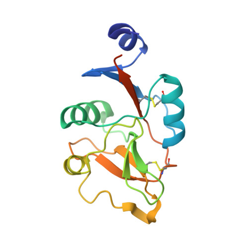
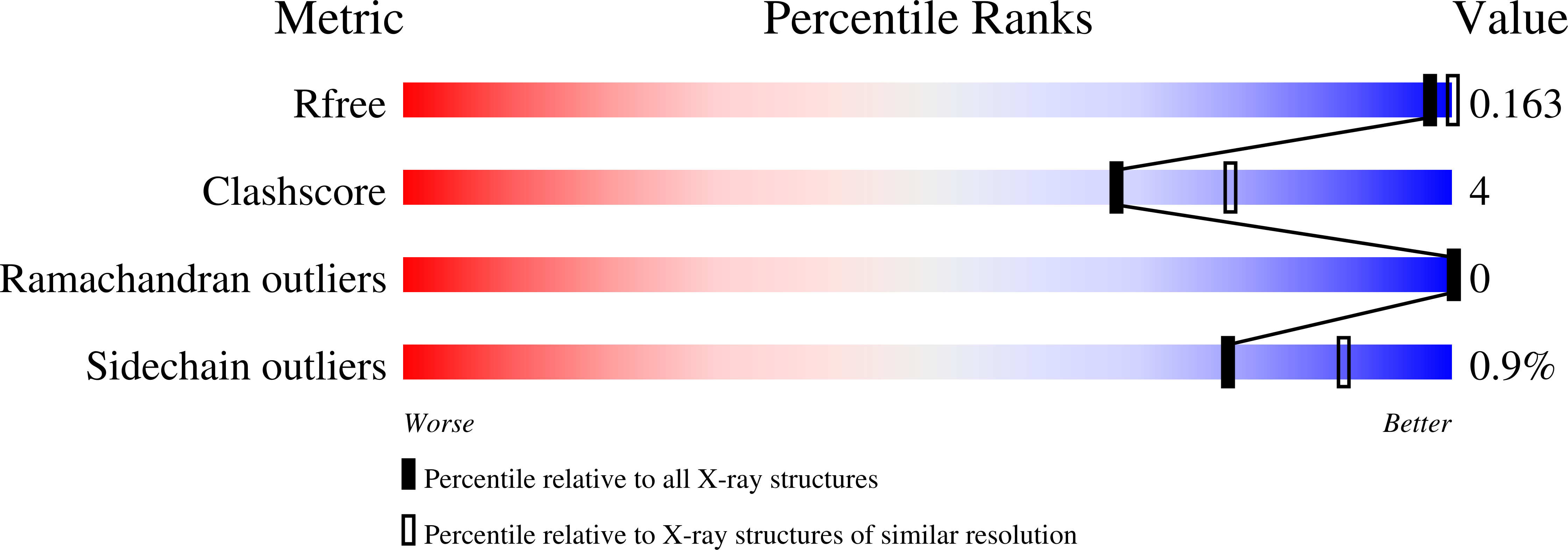
    5K8Y, 5M62

  • PubMed Abstract: 

    The recognition of pathogen surface polysaccharides by glycan-binding proteins is a cornerstone of innate host defense. Many members of the C-type lectin receptor family serve as pattern recognition receptors facilitating pathogen uptake, antigen processing, and immunomodulation. Despite the high evolutionary pressure in host-pathogen interactions, it is still widely assumed that genetic homology conveys similar specificities. Here, we investigate the ligand specificities of the human and murine forms of the myeloid C-type lectin receptor langerin for simple and complex ligands augmented by structural insight into murine langerin. Although the two homologs share the same three-dimensional structure and recognize simple ligands identically, a screening of more than 300 bacterial polysaccharides revealed highly diverging avidity and selectivity for larger and more complex glycans. Structural and evolutionary conservation analysis identified a highly variable surface adjacent to the canonic binding site, potentially forming a secondary site of interaction for large glycans.


  • Organizational Affiliation

    From the Department of Biomolecular Systems, Max Planck Institute of Colloids and Interfaces, Potsdam 14424, Germany.


Macromolecules
Find similar proteins by:  (by identity cutoff)  |  3D Structure
Entity ID: 1
MoleculeChains Sequence LengthOrganismDetailsImage
C-type lectin domain family 4 member K
A, B
151Mus musculusMutation(s): 0 
Gene Names: Cd207Clec4k
UniProt
Find proteins for Q8VBX4 (Mus musculus)
Explore Q8VBX4 
Go to UniProtKB:  Q8VBX4
Entity Groups  
Sequence Clusters30% Identity50% Identity70% Identity90% Identity95% Identity100% Identity
UniProt GroupQ8VBX4
Sequence Annotations
Expand
  • Reference Sequence
Experimental Data & Validation

Experimental Data

  • Method: X-RAY DIFFRACTION
  • Resolution: 2.40 Å
  • R-Value Free: 0.234 
  • R-Value Work: 0.196 
  • R-Value Observed: 0.198 
  • Space Group: I 2 3
Unit Cell:
Length ( Å )Angle ( ˚ )
a = 143.31α = 90
b = 143.31β = 90
c = 143.31γ = 90
Software Package:
Software NamePurpose
REFMACrefinement
XDSdata reduction
XSCALEdata scaling
PHASERphasing

Structure Validation

View Full Validation Report


Entry History 

Deposition Data

Revision History  (Full details and data files)

  • Version 1.0: 2016-12-07
    Type: Initial release
  • Version 1.1: 2016-12-14
    Changes: Database references
  • Version 1.2: 2017-02-01
    Changes: Database references
  • Version 1.3: 2024-01-10
    Changes: Data collection, Database references, Derived calculations, Refinement description