5J6H

Recognition of the MHC class Ib molecule H2-Q10 by the natural killer cell receptor Ly49C


Experimental Data Snapshot

  • Method: X-RAY DIFFRACTION
  • Resolution: 2.30 Å
  • R-Value Free: 0.243 
  • R-Value Work: 0.177 
  • R-Value Observed: 0.180 

Starting Model: experimental
View more details

wwPDB Validation   3D Report Full Report


This is version 1.6 of the entry. See complete history


Literature

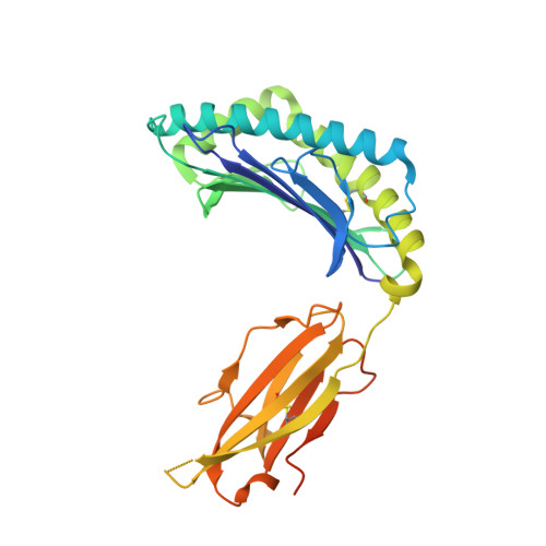
Recognition of the Major Histocompatibility Complex (MHC) Class Ib Molecule H2-Q10 by the Natural Killer Cell Receptor Ly49C.

Sullivan, L.C.Berry, R.Sosnin, N.Widjaja, J.M.Deuss, F.A.Balaji, G.R.LaGruta, N.L.Mirams, M.Trapani, J.A.Rossjohn, J.Brooks, A.G.Andrews, D.M.

(2016) J Biol Chem 291: 18740-18752

  • DOI: https://doi.org/10.1074/jbc.M116.737130
  • Primary Citation of Related Structures:  
    5J6G, 5J6H

  • PubMed Abstract: 

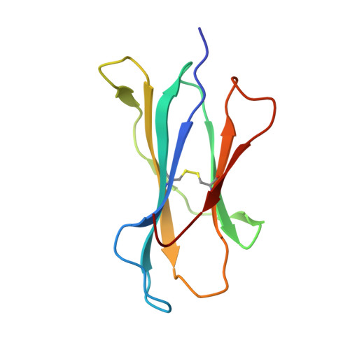
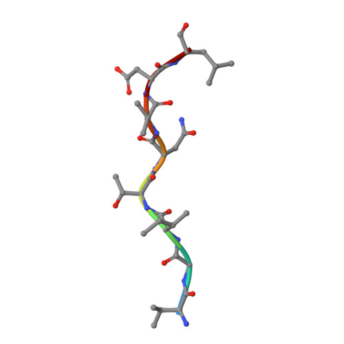
    Murine natural killer (NK) cells are regulated by the interaction of Ly49 receptors with major histocompatibility complex class I molecules (MHC-I). Although the ligands for inhibitory Ly49 were considered to be restricted to classical MHC (MHC-Ia), we have shown that the non-classical MHC molecule (MHC-Ib) H2-M3 was a ligand for the inhibitory Ly49A. Here we establish that another MHC-Ib, H2-Q10, is a bona fide ligand for the inhibitory Ly49C receptor. H2-Q10 bound to Ly49C with a marginally lower affinity (∼5 μm) than that observed between Ly49C and MHC-Ia (H-2K(b)/H-2D(d), both ∼1 μm), and this recognition could be prevented by cis interactions with H-2K in situ To understand the molecular details underpinning Ly49·MHC-Ib recognition, we determined the crystal structures of H2-Q10 and Ly49C bound H2-Q10. Unliganded H2-Q10 adopted a classical MHC-I fold and possessed a peptide-binding groove that exhibited features similar to those found in MHC-Ia, explaining the diverse peptide binding repertoire of H2-Q10. Ly49C bound to H2-Q10 underneath the peptide binding platform to a region that encompassed residues from the α1, α2, and α3 domains, as well as the associated β2-microglobulin subunit. This docking mode was conserved with that previously observed for Ly49C·H-2K(b) Indeed, structure-guided mutation of Ly49C indicated that Ly49C·H2-Q10 and Ly49C·H-2K(b) possess similar energetic footprints focused around residues located within the Ly49C β4-stand and L5 loop, which contact the underside of the peptide-binding platform floor. Our data provide a structural basis for Ly49·MHC-Ib recognition and demonstrate that MHC-Ib represent an extended family of ligands for Ly49 molecules.


  • Organizational Affiliation

    From the Department of Microbiology and Immunology, University of Melbourne, Parkville, Victoria 3010, Australia.


Macromolecules
Find similar proteins by:  (by identity cutoff)  |  3D Structure
Entity ID: 1
MoleculeChains Sequence LengthOrganismDetailsImage
H-2 class I histocompatibility antigen, Q10 alpha chain302Mus musculusMutation(s): 0 
Gene Names: H2-Q10
UniProt & NIH Common Fund Data Resources
Find proteins for P01898 (Mus musculus)
Explore P01898 
Go to UniProtKB:  P01898
IMPC:  MGI:95929
Entity Groups  
Sequence Clusters30% Identity50% Identity70% Identity90% Identity95% Identity100% Identity
UniProt GroupP01898
Sequence Annotations
Expand
  • Reference Sequence
Find similar proteins by:  (by identity cutoff)  |  3D Structure
Entity ID: 2
MoleculeChains Sequence LengthOrganismDetailsImage
Beta-2-microglobulin100Mus musculusMutation(s): 0 
Gene Names: B2m
UniProt & NIH Common Fund Data Resources
Find proteins for P01887 (Mus musculus)
Explore P01887 
Go to UniProtKB:  P01887
IMPC:  MGI:88127
Entity Groups  
Sequence Clusters30% Identity50% Identity70% Identity90% Identity95% Identity100% Identity
UniProt GroupP01887
Sequence Annotations
Expand
  • Reference Sequence

Find similar proteins by:  Sequence   |   3D Structure  

Entity ID: 3
MoleculeChains Sequence LengthOrganismDetailsImage
VAL-GLY-ILE-THR-ASN-VAL-ASP-LEUC [auth F]8Mus musculusMutation(s): 0 
UniProt & NIH Common Fund Data Resources
Find proteins for Q9DBG6 (Mus musculus)
Explore Q9DBG6 
Go to UniProtKB:  Q9DBG6
IMPC:  MGI:98085
Entity Groups  
UniProt GroupQ9DBG6
Sequence Annotations
Expand
  • Reference Sequence
Experimental Data & Validation

Experimental Data

  • Method: X-RAY DIFFRACTION
  • Resolution: 2.30 Å
  • R-Value Free: 0.243 
  • R-Value Work: 0.177 
  • R-Value Observed: 0.180 
  • Space Group: P 21 21 21
Unit Cell:
Length ( Å )Angle ( ˚ )
a = 55.42α = 90
b = 81.66β = 90
c = 97.92γ = 90
Software Package:
Software NamePurpose
BUSTERrefinement
iMOSFLMdata reduction
SCALAdata scaling
PHASERphasing

Structure Validation

View Full Validation Report


Entry History & Funding Information

Deposition Data


Funding OrganizationLocationGrant Number
National Health and Medical Research Council (NHMRC, Australia)AustraliaGNT1109901

Revision History  (Full details and data files)

  • Version 1.0: 2016-07-13
    Type: Initial release
  • Version 1.1: 2016-07-20
    Changes: Database references
  • Version 1.2: 2016-09-14
    Changes: Database references
  • Version 1.3: 2017-08-09
    Changes: Data collection, Database references, Derived calculations
  • Version 1.4: 2017-09-20
    Changes: Author supporting evidence
  • Version 1.5: 2020-01-08
    Changes: Author supporting evidence
  • Version 1.6: 2023-09-27
    Changes: Data collection, Database references, Refinement description