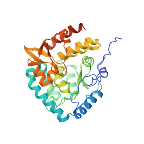
5J6D

Discovery of acyl guanidine tryptophan hydroxylase-1 inhibitors


Experimental Data Snapshot

  • Method: X-RAY DIFFRACTION
  • Resolution: 1.90 Å
  • R-Value Free: 0.248 
  • R-Value Work: 0.210 
  • R-Value Observed: 0.212 

Starting Model: experimental
View more details

wwPDB Validation   3D Report Full Report


Ligand Structure Quality Assessment 


This is version 1.2 of the entry. See complete history


Literature

Discovery of acyl guanidine tryptophan hydroxylase-1 inhibitors.

Goldberg, D.R.De Lombaert, S.Aiello, R.Bourassa, P.Barucci, N.Zhang, Q.Paralkar, V.Stein, A.J.Valentine, J.Zavadoski, W.

(2016) Bioorg Med Chem Lett 26: 2855-2860

  • DOI: https://doi.org/10.1016/j.bmcl.2016.04.057
  • Primary Citation of Related Structures:  
    5J6D

  • PubMed Abstract: 

    An increasing number of diseases have been linked to a dysfunctional peripheral serotonin system. Given that tryptophan hydroxylase 1 (TPH1) is the rate limiting enzyme in the biosynthesis off serotonin, it represents an attractive target to regulate peripheral serotonin. Following up to our first disclosure, we report a new chemotype of TPH1 inhibitors where-by the more common central planar heterocycle has been replaced with an open-chain, acyl guanidine surrogate. Through our work, we found that compounds of this nature provide highly potent TPH1 inhibitors with favorable physicochemical properties that were effective in reducing murine intestinal 5-HT in vivo. Furthermore, we obtained a high resolution (1.90Å) X-ray structure crystal structure of one of these inhibitors (compound 51) that elucidated the active conformation along with revealing a dimeric form of TPH1 for the first time.


  • Organizational Affiliation

    Karos Pharmaceuticals, 401 Winchester Ave., 5 Science Park, New Haven, CT 06511, United States. Electronic address: dgoldberg@kineta.us.


Macromolecules
Find similar proteins by:  (by identity cutoff)  |  3D Structure
Entity ID: 1
MoleculeChains Sequence LengthOrganismDetailsImage
Tryptophan 5-hydroxylase 1
A, B
307Homo sapiensMutation(s): 0 
Gene Names: TPH1TPHTPRHTRPH
EC: 1.14.16.4
UniProt & NIH Common Fund Data Resources
Find proteins for P17752 (Homo sapiens)
Explore P17752 
Go to UniProtKB:  P17752
GTEx:  ENSG00000129167 
Entity Groups  
Sequence Clusters30% Identity50% Identity70% Identity90% Identity95% Identity100% Identity
UniProt GroupP17752
Sequence Annotations
Expand
  • Reference Sequence
Small Molecules
Ligands 4 Unique
IDChains Name / Formula / InChI Key2D Diagram3D Interactions
6H5
Query on 6H5

Download Ideal Coordinates CCD File 
F [auth A],
J [auth B]
4-[(N-{[2-(3-methoxyphenoxy)-6-(piperidin-1-yl)phenyl]methyl}carbamimidoyl)carbamoyl]-L-phenylalanine
C30 H35 N5 O5
UDMPHIGEFLMAIW-VWLOTQADSA-N
TRS
Query on TRS

Download Ideal Coordinates CCD File 
D [auth A],
H [auth B]
2-AMINO-2-HYDROXYMETHYL-PROPANE-1,3-DIOL
C4 H12 N O3
LENZDBCJOHFCAS-UHFFFAOYSA-O
GOL
Query on GOL

Download Ideal Coordinates CCD File 
E [auth A],
I [auth B]
GLYCEROL
C3 H8 O3
PEDCQBHIVMGVHV-UHFFFAOYSA-N
FE
Query on FE

Download Ideal Coordinates CCD File 
C [auth A],
G [auth B]
FE (III) ION
Fe
VTLYFUHAOXGGBS-UHFFFAOYSA-N
Experimental Data & Validation

Experimental Data

  • Method: X-RAY DIFFRACTION
  • Resolution: 1.90 Å
  • R-Value Free: 0.248 
  • R-Value Work: 0.210 
  • R-Value Observed: 0.212 
  • Space Group: P 21 21 21
Unit Cell:
Length ( Å )Angle ( ˚ )
a = 58.726α = 90
b = 63.561β = 90
c = 156.468γ = 90
Software Package:
Software NamePurpose
REFMACrefinement
HKL-2000data reduction
HKL-2000data scaling
PHASERphasing

Structure Validation

View Full Validation Report


Ligand Structure Quality Assessment 


Entry History 

Deposition Data

Revision History  (Full details and data files)

  • Version 1.0: 2016-05-25
    Type: Initial release
  • Version 1.1: 2016-06-15
    Changes: Database references
  • Version 1.2: 2023-09-27
    Changes: Data collection, Database references, Derived calculations, Refinement description