Comparative Structural and Functional Analysis of Bunyavirus and Arenavirus Cap-Snatching Endonucleases.
Reguera, J., Gerlach, P., Rosenthal, M., Gaudon, S., Coscia, F., Gunther, S., Cusack, S.(2016) PLoS Pathog 12: e1005636-e1005636
- PubMed: 27304209
- DOI: https://doi.org/10.1371/journal.ppat.1005636
- Primary Citation of Related Structures:
5IZE, 5IZH, 5J1N, 5J1P - PubMed Abstract:
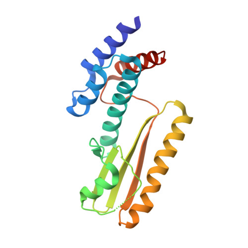
Segmented negative strand RNA viruses of the arena-, bunya- and orthomyxovirus families uniquely carry out viral mRNA transcription by the cap-snatching mechanism. This involves cleavage of host mRNAs close to their capped 5' end by an endonuclease (EN) domain located in the N-terminal region of the viral polymerase. We present the structure of the cap-snatching EN of Hantaan virus, a bunyavirus belonging to hantavirus genus. Hantaan EN has an active site configuration, including a metal co-ordinating histidine, and nuclease activity similar to the previously reported La Crosse virus and Influenza virus ENs (orthobunyavirus and orthomyxovirus respectively), but is more active in cleaving a double stranded RNA substrate. In contrast, Lassa arenavirus EN has only acidic metal co-ordinating residues. We present three high resolution structures of Lassa virus EN with different bound ion configurations and show in comparative biophysical and biochemical experiments with Hantaan, La Crosse and influenza ENs that the isolated Lassa EN is essentially inactive. The results are discussed in the light of EN activation mechanisms revealed by recent structures of full-length influenza virus polymerase.
Organizational Affiliation:
European Molecular Biology Laboratory, Grenoble Outstation, 71 Avenue des Martyrs, CS90181, 38042 Grenoble Cedex 9, France.


