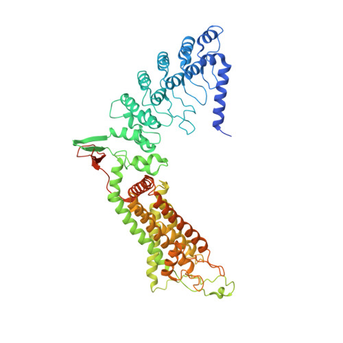
5IWT

Structure of Transient Receptor Potential (TRP) channel TRPV6 in the presence of gadolinium


Experimental Data Snapshot

  • Method: X-RAY DIFFRACTION
  • Resolution: 3.80 Å
  • R-Value Free: 0.307 
  • R-Value Work: 0.275 
  • R-Value Observed: 0.277 

Starting Model: experimental
View more details

wwPDB Validation   3D Report Full Report


Ligand Structure Quality Assessment 


This is version 1.5 of the entry. See complete history


Literature

Crystal structure of the epithelial calcium channel TRPV6.

Saotome, K.Singh, A.K.Yelshanskaya, M.V.Sobolevsky, A.I.

(2016) Nature 534: 506-511

  • DOI: https://doi.org/10.1038/nature17975
  • Primary Citation of Related Structures:  
    5IWK, 5IWP, 5IWR, 5IWT

  • PubMed Abstract: 

    Precise regulation of calcium homeostasis is essential for many physiological functions. The Ca(2+)-selective transient receptor potential (TRP) channels TRPV5 and TRPV6 play vital roles in calcium homeostasis as Ca(2+) uptake channels in epithelial tissues. Detailed structural bases for their assembly and Ca(2+) permeation remain obscure. Here we report the crystal structure of rat TRPV6 at 3.25 Å resolution. The overall architecture of TRPV6 reveals shared and unique features compared with other TRP channels. Intracellular domains engage in extensive interactions to form an intracellular 'skirt' involved in allosteric modulation. In the K(+) channel-like transmembrane domain, Ca(2+) selectivity is determined by direct coordination of Ca(2+) by a ring of aspartate side chains in the selectivity filter. On the basis of crystallographically identified cation-binding sites at the pore axis and extracellular vestibule, we propose a Ca(2+) permeation mechanism. Our results provide a structural foundation for understanding the regulation of epithelial Ca(2+) uptake and its role in pathophysiology.


  • Organizational Affiliation

    Department of Biochemistry and Molecular Biophysics, Columbia University, 650 West 168th Street, New York, New York 10032, USA.


Macromolecules
Find similar proteins by:  (by identity cutoff)  |  3D Structure
Entity ID: 1
MoleculeChains Sequence LengthOrganismDetailsImage
Transient receptor potential cation channel subfamily V member 6672Rattus norvegicusMutation(s): 4 
Gene Names: Trpv6
Membrane Entity: Yes 
UniProt
Find proteins for Q9R186 (Rattus norvegicus)
Explore Q9R186 
Go to UniProtKB:  Q9R186
Entity Groups  
Sequence Clusters30% Identity50% Identity70% Identity90% Identity95% Identity100% Identity
UniProt GroupQ9R186
Sequence Annotations
Expand
  • Reference Sequence
Small Molecules
Experimental Data & Validation

Experimental Data

  • Method: X-RAY DIFFRACTION
  • Resolution: 3.80 Å
  • R-Value Free: 0.307 
  • R-Value Work: 0.275 
  • R-Value Observed: 0.277 
  • Space Group: P 4 21 2
Unit Cell:
Length ( Å )Angle ( ˚ )
a = 144.35α = 90
b = 144.35β = 90
c = 113.37γ = 90
Software Package:
Software NamePurpose
PHENIXrefinement
XDSdata reduction
XDSdata scaling
PHASERphasing

Structure Validation

View Full Validation Report


Ligand Structure Quality Assessment 


Entry History & Funding Information

Deposition Data


Funding OrganizationLocationGrant Number
National Institutes of Health/National Institute of Neurological Disorders and Stroke (NIH/NINDS)United StatesR01 NS083660
National Institutes of Health/National Institute of General Medical Sciences (NIH/NIGMS)United StatesT32 GM008281

Revision History  (Full details and data files)

  • Version 1.0: 2016-06-15
    Type: Initial release
  • Version 1.1: 2016-06-29
    Changes: Database references
  • Version 1.2: 2016-07-06
    Changes: Data collection
  • Version 1.3: 2017-09-20
    Changes: Author supporting evidence, Derived calculations
  • Version 1.4: 2019-12-18
    Changes: Author supporting evidence
  • Version 1.5: 2023-09-27
    Changes: Data collection, Database references, Refinement description