5IQU

WelO5 G166D variant bound to Fe(II), 2-oxoglutarate, and 12-epifischerindole U


Experimental Data Snapshot

  • Method: X-RAY DIFFRACTION
  • Resolution: 2.51 Å
  • R-Value Free: 0.262 
  • R-Value Work: 0.223 
  • R-Value Observed: 0.225 

Starting Model: experimental
View more details

wwPDB Validation   3D Report Full Report


Ligand Structure Quality Assessment 


This is version 1.5 of the entry. See complete history


Literature

Structural basis for halogenation by iron- and 2-oxo-glutarate-dependent enzyme WelO5.

Mitchell, A.J.Zhu, Q.Maggiolo, A.O.Ananth, N.R.Hillwig, M.L.Liu, X.Boal, A.K.

(2016) Nat Chem Biol 12: 636-640

  • DOI: https://doi.org/10.1038/nchembio.2112
  • Primary Citation of Related Structures:  
    5IQS, 5IQT, 5IQU, 5IQV

  • PubMed Abstract: 

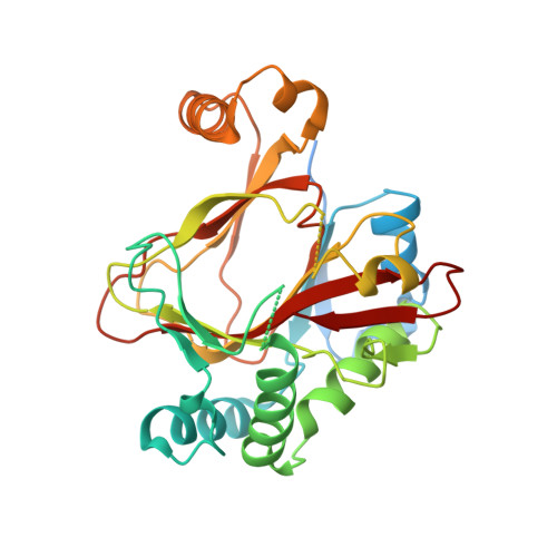
    A 2.4-Å-resolution X-ray crystal structure of the carrier-protein-independent halogenase WelO5 in complex with its welwitindolinone precursor substrate, 12-epi-fischerindole U, reveals that the C13 chlorination target is proximal to the anticipated site of the oxo group in a presumptive cis-halo-oxo-iron(IV) (haloferryl) intermediate. Prior study of related halogenases forecasts substrate hydroxylation in this active-site configuration, but X-ray crystallographic verification of C13 halogenation in single crystals mandates that ligand dynamics must reposition the oxygen ligand to enable the observed outcome. S189A WelO5 produces a mixture of halogenation and hydroxylation products, showing that an outer-sphere hydrogen-bonding group orchestrates ligand movements to achieve a configuration that promotes halogen transfer.


  • Organizational Affiliation

    Department of Biochemistry and Molecular Biology, The Pennsylvania State University, University Park, Pennsylvania, USA.


Macromolecules
Find similar proteins by:  (by identity cutoff)  |  3D Structure
Entity ID: 1
MoleculeChains Sequence LengthOrganismDetailsImage
WelO5315Hapalosiphon welwitschii UTEX B 1830Mutation(s): 1 
UniProt
Find proteins for A0A067YX61 (Hapalosiphon welwitschii UTEX B 1830)
Explore A0A067YX61 
Go to UniProtKB:  A0A067YX61
Entity Groups  
Sequence Clusters30% Identity50% Identity70% Identity90% Identity95% Identity100% Identity
UniProt GroupA0A067YX61
Sequence Annotations
Expand
  • Reference Sequence
Small Molecules
Ligands 3 Unique
IDChains Name / Formula / InChI Key2D Diagram3D Interactions
6CU
Query on 6CU

Download Ideal Coordinates CCD File 
D [auth A](6aS,9R,10R,10aS)-9-ethyl-10-isocyano-6,6,9-trimethyl-5,6,6a,7,8,9,10,10a-octahydroindeno[2,1-b]indole
C21 H26 N2
BWQHHWNHLMXCLS-QBGRRASTSA-N
AKG
Query on AKG

Download Ideal Coordinates CCD File 
C [auth A]2-OXOGLUTARIC ACID
C5 H6 O5
KPGXRSRHYNQIFN-UHFFFAOYSA-N
FE2
Query on FE2

Download Ideal Coordinates CCD File 
B [auth A]FE (II) ION
Fe
CWYNVVGOOAEACU-UHFFFAOYSA-N
Experimental Data & Validation

Experimental Data

  • Method: X-RAY DIFFRACTION
  • Resolution: 2.51 Å
  • R-Value Free: 0.262 
  • R-Value Work: 0.223 
  • R-Value Observed: 0.225 
  • Space Group: C 1 2 1
Unit Cell:
Length ( Å )Angle ( ˚ )
a = 74.107α = 90
b = 54.127β = 90.79
c = 74.639γ = 90
Software Package:
Software NamePurpose
HKL-2000data scaling
REFMACrefinement
PDB_EXTRACTdata extraction
PHASERphasing
HKL-2000data reduction

Structure Validation

View Full Validation Report


Ligand Structure Quality Assessment 


Entry History & Funding Information

Deposition Data


Funding OrganizationLocationGrant Number
National Institutes of Health/National Institute of General Medical Sciences (NIH/NIGMS)United StatesGM100011

Revision History  (Full details and data files)

  • Version 1.0: 2016-06-29
    Type: Initial release
  • Version 1.1: 2016-07-13
    Changes: Database references
  • Version 1.2: 2016-07-27
    Changes: Database references
  • Version 1.3: 2017-09-13
    Changes: Author supporting evidence, Derived calculations
  • Version 1.4: 2022-04-13
    Changes: Author supporting evidence, Database references, Derived calculations
  • Version 1.5: 2023-09-27
    Changes: Data collection, Refinement description