


Crystal Structure of the alpha spectrin SH3 domain mutant N47A
Camara-Artigas, A.To be published.
Experimental Data Snapshot
Starting Model: experimental
View more details
wwPDB Validation 3D Report Full Report

Entity ID: 1 | |||||
|---|---|---|---|---|---|
| Molecule | Chains | Sequence Length | Organism | Details | Image |
| Spectrin alpha chain, non-erythrocytic 1 | 62 | Gallus gallus | Mutation(s): 1 Gene Names: SPTAN1, SPTA2 | 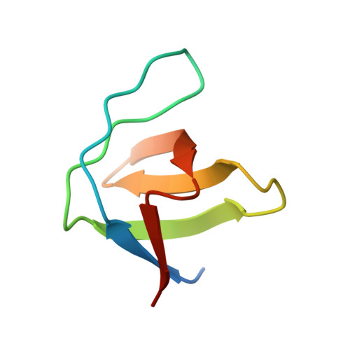 | |
UniProt | |||||
Find proteins for P07751 (Gallus gallus) Explore P07751 Go to UniProtKB: P07751 | |||||
Entity Groups | |||||
| Sequence Clusters | 30% Identity50% Identity70% Identity90% Identity95% Identity100% Identity | ||||
| UniProt Group | P07751 | ||||
Sequence AnnotationsExpand | |||||
| |||||
| Ligands 3 Unique | |||||
|---|---|---|---|---|---|
| ID | Chains | Name / Formula / InChI Key | 2D Diagram | 3D Interactions | |
| PRO Query on PRO | C [auth A] | PROLINE C5 H9 N O2 ONIBWKKTOPOVIA-BYPYZUCNSA-N |  | ||
| SO4 Query on SO4 | B [auth A] | SULFATE ION O4 S QAOWNCQODCNURD-UHFFFAOYSA-L |  | ||
| ACT Query on ACT | D [auth A] | ACETATE ION C2 H3 O2 QTBSBXVTEAMEQO-UHFFFAOYSA-M |  | ||
| Length ( Å ) | Angle ( ˚ ) |
|---|---|
| a = 33.55 | α = 90 |
| b = 42.371 | β = 90 |
| c = 49.544 | γ = 90 |
| Software Name | Purpose |
|---|---|
| Aimless | data scaling |
| PHASER | phasing |
| PHENIX | refinement |
| PDB_EXTRACT | data extraction |
| XDS | data reduction |
RCSB PDB (citation) is hosted by
RCSB PDB is a member of the

