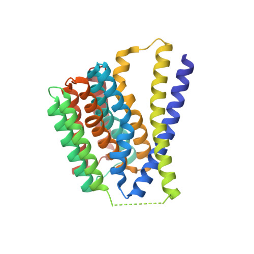
5HYA

Structural mechanisms of extracellular ion exchange and induced binding-site occlusion in the sodium-calcium exchangerNCX_Mj soaked with 150 mM Na+ and nominal Ca2+


Experimental Data Snapshot

  • Method: X-RAY DIFFRACTION
  • Resolution: 1.90 Å
  • R-Value Free: 0.207 
  • R-Value Work: 0.179 
  • R-Value Observed: 0.181 

Starting Model: experimental
View more details

wwPDB Validation   3D Report Full Report


Ligand Structure Quality Assessment 


This is version 1.4 of the entry. See complete history


Literature

Mechanism of extracellular ion exchange and binding-site occlusion in a sodium/calcium exchanger

Liao, J.Marinelli, F.Lee, C.Huang, Y.Faraldo-Gomez, J.D.Jiang, Y.

(2016) Nat Struct Mol Biol 23: 590-599

  • DOI: https://doi.org/10.1038/nsmb.3230
  • Primary Citation of Related Structures:  
    5HWX, 5HWY, 5HXC, 5HXE, 5HXH, 5HXR, 5HXS, 5HYA, 5JDF, 5JDG, 5JDH, 5JDL, 5JDM, 5JDN, 5JDQ

  • PubMed Abstract: 

    Na(+)/Ca(2+) exchangers use the Na(+) electrochemical gradient across the plasma membrane to extrude intracellular Ca(2+) and play a central role in Ca(2+) homeostasis. Here, we elucidate their mechanisms of extracellular ion recognition and exchange through a structural analysis of the exchanger from Methanococcus jannaschii (NCX_Mj) bound to Na(+), Ca(2+) or Sr(2+) in various occupancies and in an apo state. This analysis defines the binding mode and relative affinity of these ions, establishes the structural basis for the anticipated 3:1 Na(+)/Ca(2+)-exchange stoichiometry and reveals the conformational changes at the onset of the alternating-access transport mechanism. An independent analysis of the dynamics and conformational free-energy landscape of NCX_Mj in different ion-occupancy states, based on enhanced-sampling molecular dynamics simulations, demonstrates that the crystal structures reflect mechanistically relevant, interconverting conformations. These calculations also reveal the mechanism by which the outward-to-inward transition is controlled by the ion occupancy, thereby explaining the emergence of strictly coupled Na(+)/Ca(2+) antiport.


  • Organizational Affiliation

    School of Life Science and Technology, ShanghaiTech University, Shanghai, P.R. China.


Macromolecules
Find similar proteins by:  (by identity cutoff)  |  3D Structure
Entity ID: 1
MoleculeChains Sequence LengthOrganismDetailsImage
sodium,calcium exchanger304Methanocaldococcus jannaschii DSM 2661Mutation(s): 1 
Gene Names: MJ0091
Membrane Entity: Yes 
UniProt
Find proteins for Q57556 (Methanocaldococcus jannaschii (strain ATCC 43067 / DSM 2661 / JAL-1 / JCM 10045 / NBRC 100440))
Explore Q57556 
Go to UniProtKB:  Q57556
Entity Groups  
Sequence Clusters30% Identity50% Identity70% Identity90% Identity95% Identity100% Identity
UniProt GroupQ57556
Sequence Annotations
Expand
  • Reference Sequence
Small Molecules
Ligands 6 Unique
IDChains Name / Formula / InChI Key2D Diagram3D Interactions
OLC
Query on OLC

Download Ideal Coordinates CCD File 
J [auth A],
K [auth A],
L [auth A],
W [auth A],
X [auth A]
(2R)-2,3-dihydroxypropyl (9Z)-octadec-9-enoate
C21 H40 O4
RZRNAYUHWVFMIP-GDCKJWNLSA-N
1PE
Query on 1PE

Download Ideal Coordinates CCD File 
P [auth A]PENTAETHYLENE GLYCOL
C10 H22 O6
JLFNLZLINWHATN-UHFFFAOYSA-N
MYS
Query on MYS

Download Ideal Coordinates CCD File 
M [auth A]
N [auth A]
O [auth A]
Q [auth A]
R [auth A]
M [auth A],
N [auth A],
O [auth A],
Q [auth A],
R [auth A],
S [auth A],
T [auth A],
U [auth A],
V [auth A]
PENTADECANE
C15 H32
YCOZIPAWZNQLMR-UHFFFAOYSA-N
ACT
Query on ACT

Download Ideal Coordinates CCD File 
F [auth A],
G [auth A],
H [auth A],
I [auth A]
ACETATE ION
C2 H3 O2
QTBSBXVTEAMEQO-UHFFFAOYSA-M
CA
Query on CA

Download Ideal Coordinates CCD File 
E [auth A]CALCIUM ION
Ca
BHPQYMZQTOCNFJ-UHFFFAOYSA-N
NA
Query on NA

Download Ideal Coordinates CCD File 
B [auth A],
C [auth A],
D [auth A]
SODIUM ION
Na
FKNQFGJONOIPTF-UHFFFAOYSA-N
Experimental Data & Validation

Experimental Data

  • Method: X-RAY DIFFRACTION
  • Resolution: 1.90 Å
  • R-Value Free: 0.207 
  • R-Value Work: 0.179 
  • R-Value Observed: 0.181 
  • Space Group: P 21 21 21
Unit Cell:
Length ( Å )Angle ( ˚ )
a = 49.49α = 90
b = 72.879β = 90
c = 96.206γ = 90
Software Package:
Software NamePurpose
PHENIXrefinement
HKL-2000data scaling
PDB_EXTRACTdata extraction
HKL-2000data reduction
SHARPphasing

Structure Validation

View Full Validation Report


Ligand Structure Quality Assessment 


Entry History & Funding Information

Deposition Data


Funding OrganizationLocationGrant Number
National Natural Science Foundation of ChinaChinaProject 31470817
Howard Hughes Medical InstituteUnited StatesFor Youxing Jiang
National Institutes of Health/National Institute of General Medical Sciences (NIH/NIGMS)United StatesR01GM079179
Welch FoundationUnited StatesGrant I-1578
National Institutes of HealthUnited StatesDivision of Intramural Research of the National Heart, Lung and Blood Institute

Revision History  (Full details and data files)

  • Version 1.0: 2016-05-11
    Type: Initial release
  • Version 1.1: 2016-06-22
    Changes: Database references
  • Version 1.2: 2017-10-18
    Changes: Author supporting evidence, Derived calculations, Structure summary
  • Version 1.3: 2022-03-23
    Changes: Author supporting evidence, Database references, Derived calculations
  • Version 1.4: 2023-11-08
    Changes: Data collection, Refinement description