5HIX

Cocrystal structure of an anti-parallel DNA G-quadruplex and a tetra-Quinoline Foldamer


Experimental Data Snapshot

  • Method: X-RAY DIFFRACTION
  • Resolution: 2.48 Å
  • R-Value Free: 0.288 
  • R-Value Work: 0.227 
  • R-Value Observed: 0.231 

Starting Model: experimental
View more details

wwPDB Validation   3D Report Full Report


Ligand Structure Quality Assessment 


This is version 1.1 of the entry. See complete history


Literature

Multivalent Interactions between an Aromatic Helical Foldamer and a DNA G-Quadruplex in the Solid State.

Mandal, P.K.Baptiste, B.Langlois d'Estaintot, B.Kauffmann, B.Huc, I.

(2016) Chembiochem 17: 1911-1914

  • DOI: https://doi.org/10.1002/cbic.201600281
  • Primary Citation of Related Structures:  
    5HIX

  • PubMed Abstract: 

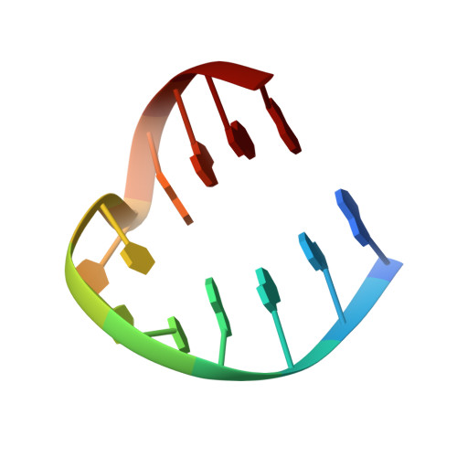
    Quinoline-based oligoamide foldamers have been identified as a potent class of ligands for G-quadruplex DNA. Their helical structure is thought to target G-quadruplex loops or grooves and not G-tetrads. We report a co-crystal structure of the antiparallel hairpin dimeric DNA G-quadruplex (G 4 T 4 G 4 ) 2 with tetramer 1-a helically folded oligo-quinolinecarboxamide bearing cationic side chains-that is consistent with this hypothesis. Multivalent foldamer-DNA interactions that modify the packing of (G 4 T 4 G 4 ) 2 in the solid state are observed.


  • Organizational Affiliation

    University of Bordeaux, CBMN, UMR 5248), Institut Européen de Chimie et Biologie, 2 rue Escarpit, 33600, Pessac, France.


Macromolecules

Find similar nucleic acids by:  Sequence   |   3D Structure  

Entity ID: 1
MoleculeChains LengthOrganismImage
Dimeric G-quadruplex
A, B
12synthetic construct
Sequence Annotations
Expand
  • Reference Sequence
Small Molecules
Ligands 2 Unique
IDChains Name / Formula / InChI Key2D Diagram3D Interactions
61Q
Query on 61Q

Download Ideal Coordinates CCD File 
G [auth A]4-(3-aminopropoxy)-8-({[4-(3-aminopropoxy)-8-({[4-(3-aminopropoxy)-8-({[4-(3-aminopropoxy)-8-nitroquinolin-2-yl]carbonyl}amino)quinolin-2-yl]carbonyl}amino)quinolin-2-yl]carbonyl}amino)quinoline-2-carboxylic acid
C52 H52 N12 O11
PDPDTUNURBJNEI-UHFFFAOYSA-N
K
Query on K

Download Ideal Coordinates CCD File 
C [auth A],
D [auth A],
E [auth A],
F [auth A]
POTASSIUM ION
K
NPYPAHLBTDXSSS-UHFFFAOYSA-N
Experimental Data & Validation

Experimental Data

  • Method: X-RAY DIFFRACTION
  • Resolution: 2.48 Å
  • R-Value Free: 0.288 
  • R-Value Work: 0.227 
  • R-Value Observed: 0.231 
  • Space Group: P 41
Unit Cell:
Length ( Å )Angle ( ˚ )
a = 31.701α = 90
b = 31.701β = 90
c = 97.373γ = 90
Software Package:
Software NamePurpose
REFMACrefinement
XDSdata reduction
XDSdata scaling
AMoREphasing

Structure Validation

View Full Validation Report


Ligand Structure Quality Assessment 


Entry History & Funding Information

Deposition Data


Funding OrganizationLocationGrant Number
European Research CouncilFranceERC-2012-AdG-320892

Revision History  (Full details and data files)

  • Version 1.0: 2016-11-02
    Type: Initial release
  • Version 1.1: 2024-01-10
    Changes: Data collection, Database references, Derived calculations, Refinement description