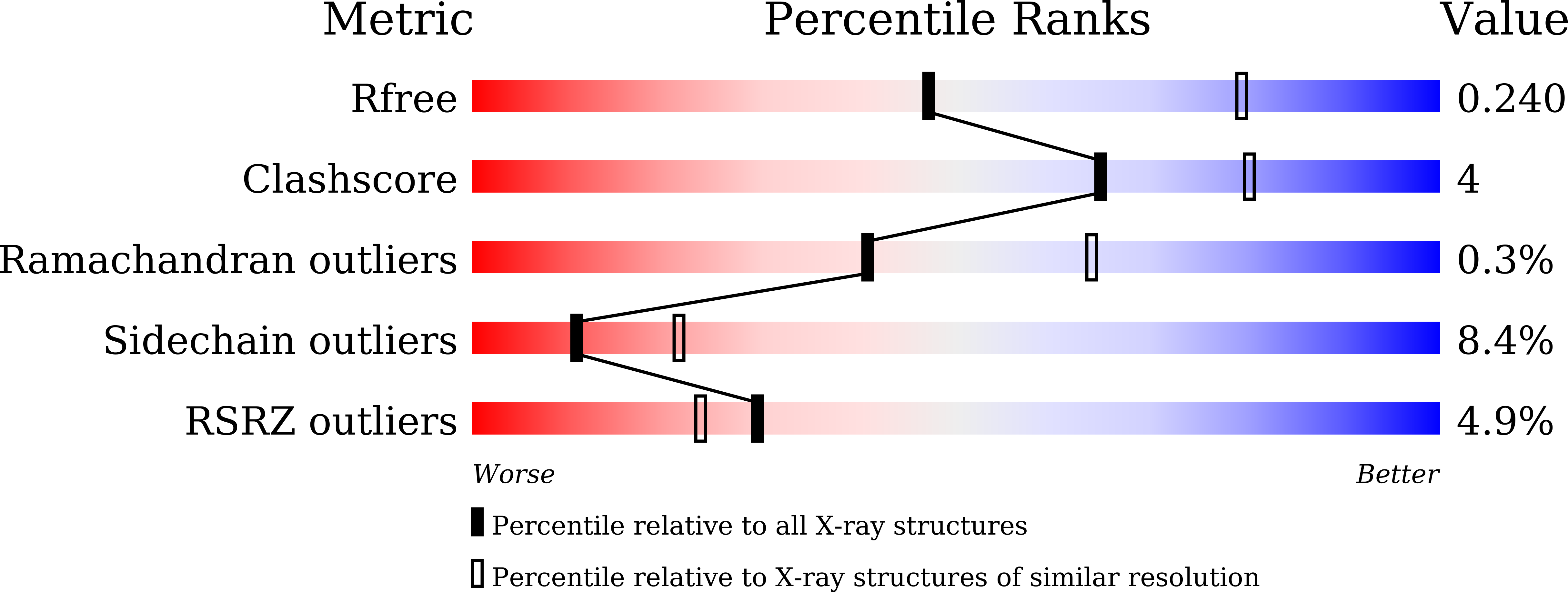
5H9W

Crystal structure of Regnase PIN domain, form II


Experimental Data Snapshot

  • Method: X-RAY DIFFRACTION
  • Resolution: 2.60 Å
  • R-Value Free: 0.231 
  • R-Value Work: 0.191 
  • R-Value Observed: 0.193 

Starting Model: experimental
View more details

wwPDB Validation   3D Report Full Report


This is version 1.2 of the entry. See complete history


Literature

Structural basis for the regulation of enzymatic activity of Regnase-1 by domain-domain interactions

Yokogawa, M.Tsushima, T.Noda, N.N.Kumeta, H.Enokizono, Y.Yamashita, K.Standley, D.M.Takeuchi, O.Akira, S.Inagaki, F.

(2016) Sci Rep 6: 22324-22324

  • DOI: https://doi.org/10.1038/srep22324
  • Primary Citation of Related Structures:  
    2N5J, 2N5K, 2N5L, 5H9V, 5H9W

  • PubMed Abstract: 

    Regnase-1 is an RNase that directly cleaves mRNAs of inflammatory genes such as IL-6 and IL-12p40, and negatively regulates cellular inflammatory responses. Here, we report the structures of four domains of Regnase-1 from Mus musculus-the N-terminal domain (NTD), PilT N-terminus like (PIN) domain, zinc finger (ZF) domain and C-terminal domain (CTD). The PIN domain harbors the RNase catalytic center; however, it is insufficient for enzymatic activity. We found that the NTD associates with the PIN domain and significantly enhances its RNase activity. The PIN domain forms a head-to-tail oligomer and the dimer interface overlaps with the NTD binding site. Interestingly, mutations blocking PIN oligomerization had no RNase activity, indicating that both oligomerization and NTD binding are crucial for RNase activity in vitro. These results suggest that Regnase-1 RNase activity is tightly controlled by both intramolecular (NTD-PIN) and intermolecular (PIN-PIN) interactions.


  • Organizational Affiliation

    Faculty of Advanced Life Science, Hokkaido University, Sapporo 001-0021, Japan.


Macromolecules
Find similar proteins by:  (by identity cutoff)  |  3D Structure
Entity ID: 1
MoleculeChains Sequence LengthOrganismDetailsImage
Ribonuclease ZC3H12A
A, B
210Mus musculusMutation(s): 0 
Gene Names: Zc3h12aMcpipMcpip1
EC: 3.1
UniProt & NIH Common Fund Data Resources
Find proteins for Q5D1E7 (Mus musculus)
Explore Q5D1E7 
Go to UniProtKB:  Q5D1E7
IMPC:  MGI:2385891
Entity Groups  
Sequence Clusters30% Identity50% Identity70% Identity90% Identity95% Identity100% Identity
UniProt GroupQ5D1E7
Sequence Annotations
Expand
  • Reference Sequence
Experimental Data & Validation

Experimental Data

  • Method: X-RAY DIFFRACTION
  • Resolution: 2.60 Å
  • R-Value Free: 0.231 
  • R-Value Work: 0.191 
  • R-Value Observed: 0.193 
  • Space Group: P 31 2 1
Unit Cell:
Length ( Å )Angle ( ˚ )
a = 113.333α = 90
b = 113.333β = 90
c = 78.373γ = 120
Software Package:
Software NamePurpose
REFMACrefinement
HKL-2000data reduction
HKL-2000data scaling
CNSphasing

Structure Validation

View Full Validation Report


Entry History & Funding Information

Deposition Data


Funding OrganizationLocationGrant Number
JSPSJapan--

Revision History  (Full details and data files)

  • Version 1.0: 2016-03-16
    Type: Initial release
  • Version 1.1: 2020-02-19
    Changes: Data collection, Derived calculations
  • Version 1.2: 2023-11-08
    Changes: Data collection, Database references, Refinement description