5FH1

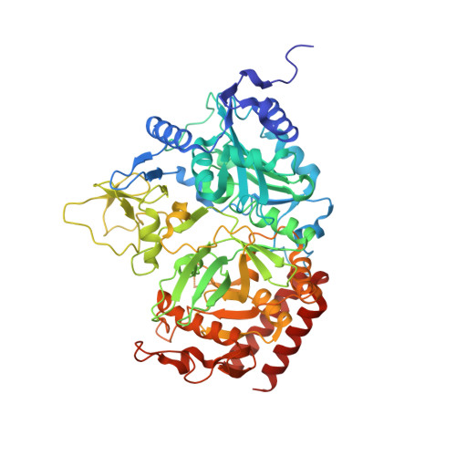
The structure of rat cytosolic PEPCK variant E89D in complex with GTP

  • Classification: LYASE
  • Organism(s): Rattus norvegicus
  • Expression System: Escherichia coli
  • Mutation(s): Yes 

  • Deposited: 2015-12-21 Released: 2016-12-28 
  • Deposition Author(s): Johnson, T.A., Holyoak, T.
  • Funding Organization(s): National Institutes of Health/National Center for Research Resources (NIH/NCRR), Natural Sciences and Engineering Research Council (NSERC, Canada)

Experimental Data Snapshot

  • Method: X-RAY DIFFRACTION
  • Resolution: 1.55 Å
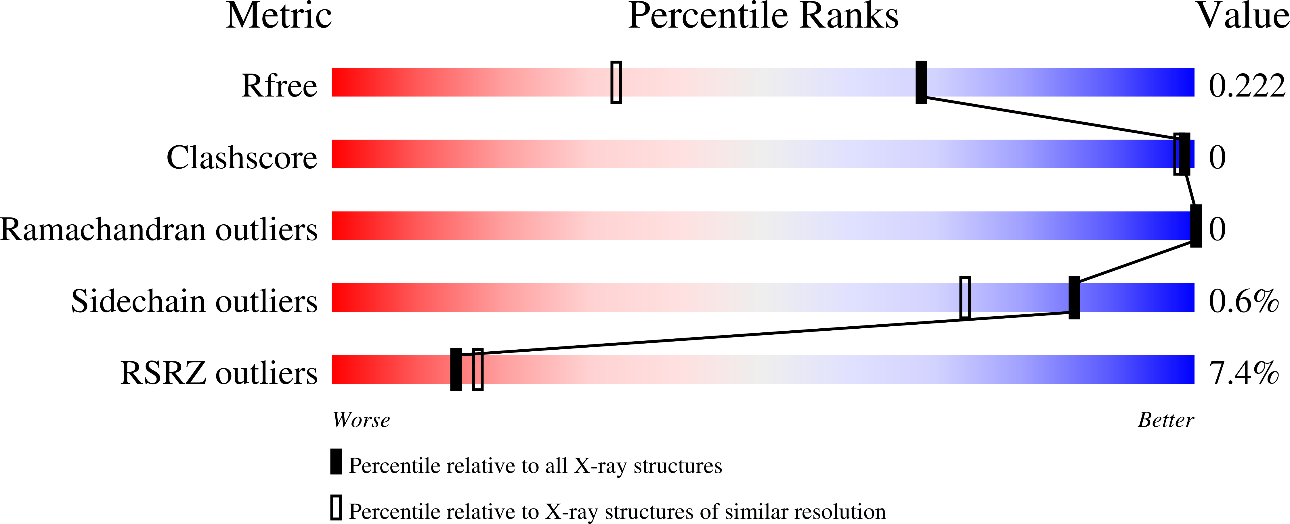
  • R-Value Free: 0.218 
  • R-Value Work: 0.183 
  • R-Value Observed: 0.185 

Starting Model: experimental
View more details

wwPDB Validation   3D Report Full Report


Ligand Structure Quality Assessment 


This is version 1.3 of the entry. See complete history


Literature

Utilization of Substrate Intrinsic Binding Energy for Conformational Change and Catalytic Function in Phosphoenolpyruvate Carboxykinase.

Johnson, T.A.Mcleod, M.J.Holyoak, T.

(2016) Biochemistry 55: 575-587

  • DOI: https://doi.org/10.1021/acs.biochem.5b01215
  • Primary Citation of Related Structures:  
    5FH0, 5FH1, 5FH2, 5FH3, 5FH4, 5FH5

  • PubMed Abstract: 

    Phosphoenolpyruvate carboxykinase (PEPCK) is an essential metabolic enzyme operating in the gluconeogenesis and glyceroneogenesis pathways. Previous work has demonstrated that the enzyme cycles between a catalytically inactive open state and a catalytically active closed state. The transition of the enzyme between these states requires the transition of several active site loops to shift from mobile, disordered structural elements to stable ordered states. The mechanism by which these disorder-order transitions are coupled to the ligation state of the active site however is not fully understood. To further investigate the mechanisms by which the mobility of the active site loops is coupled to enzymatic function and the transitioning of the enzyme between the two conformational states, we have conducted structural and functional studies of point mutants of E89. E89 is a proposed key member of the interaction network of mobile elements as it resides in the R-loop region of the enzyme active site. These new data demonstrate the importance of the R-loop in coordinating interactions between substrates at the OAA/PEP binding site and the mobile R- and Ω-loop domains. In turn, the studies more generally demonstrate the mechanisms by which the intrinsic ligand binding energy can be utilized in catalysis to drive unfavorable conformational changes, changes that are subsequently required for both optimal catalytic activity and fidelity.


  • Organizational Affiliation

    Department of Biochemistry and Molecular Biology, The University of Kansas Medical Center , Kansas City, Kansas 66160, United States.


Macromolecules
Find similar proteins by:  (by identity cutoff)  |  3D Structure
Entity ID: 1
MoleculeChains Sequence LengthOrganismDetailsImage
Phosphoenolpyruvate carboxykinase, cytosolic [GTP]622Rattus norvegicusMutation(s): 1 
Gene Names: Pck1
EC: 4.1.1.32 (PDB Primary Data), 2.7.11 (UniProt)
UniProt
Find proteins for P07379 (Rattus norvegicus)
Explore P07379 
Go to UniProtKB:  P07379
Entity Groups  
Sequence Clusters30% Identity50% Identity70% Identity90% Identity95% Identity100% Identity
UniProt GroupP07379
Sequence Annotations
Expand
  • Reference Sequence
Small Molecules
Ligands 4 Unique
IDChains Name / Formula / InChI Key2D Diagram3D Interactions
GTP
Query on GTP

Download Ideal Coordinates CCD File 
D [auth A]GUANOSINE-5'-TRIPHOSPHATE
C10 H16 N5 O14 P3
XKMLYUALXHKNFT-UUOKFMHZSA-N
EPE
Query on EPE

Download Ideal Coordinates CCD File 
E [auth A]4-(2-HYDROXYETHYL)-1-PIPERAZINE ETHANESULFONIC ACID
C8 H18 N2 O4 S
JKMHFZQWWAIEOD-UHFFFAOYSA-N
MN
Query on MN

Download Ideal Coordinates CCD File 
B [auth A],
C [auth A]
MANGANESE (II) ION
Mn
WAEMQWOKJMHJLA-UHFFFAOYSA-N
NA
Query on NA

Download Ideal Coordinates CCD File 
F [auth A]SODIUM ION
Na
FKNQFGJONOIPTF-UHFFFAOYSA-N
Experimental Data & Validation

Experimental Data

  • Method: X-RAY DIFFRACTION
  • Resolution: 1.55 Å
  • R-Value Free: 0.218 
  • R-Value Work: 0.183 
  • R-Value Observed: 0.185 
  • Space Group: P 1 21 1
Unit Cell:
Length ( Å )Angle ( ˚ )
a = 45.523α = 90
b = 119.119β = 107.66
c = 60.894γ = 90
Software Package:
Software NamePurpose
HKL-2000data collection
SCALEPACKdata scaling
REFMACrefinement
PDB_EXTRACTdata extraction
HKL-2000data reduction
MOLREPphasing

Structure Validation

View Full Validation Report


Ligand Structure Quality Assessment 


Entry History & Funding Information

Deposition Data


Funding OrganizationLocationGrant Number
National Institutes of Health/National Center for Research Resources (NIH/NCRR)United StatesP20 RR17708
Natural Sciences and Engineering Research Council (NSERC, Canada)Canada--

Revision History  (Full details and data files)

  • Version 1.0: 2016-12-28
    Type: Initial release
  • Version 1.1: 2017-09-20
    Changes: Author supporting evidence
  • Version 1.2: 2019-12-04
    Changes: Author supporting evidence
  • Version 1.3: 2023-09-27
    Changes: Data collection, Database references, Derived calculations, Refinement description