5EZ7

Crystal structure of the FAD dependent oxidoreductase PA4991 from Pseudomonas aeruginosa


Experimental Data Snapshot

  • Method: X-RAY DIFFRACTION
  • Resolution: 2.40 Å
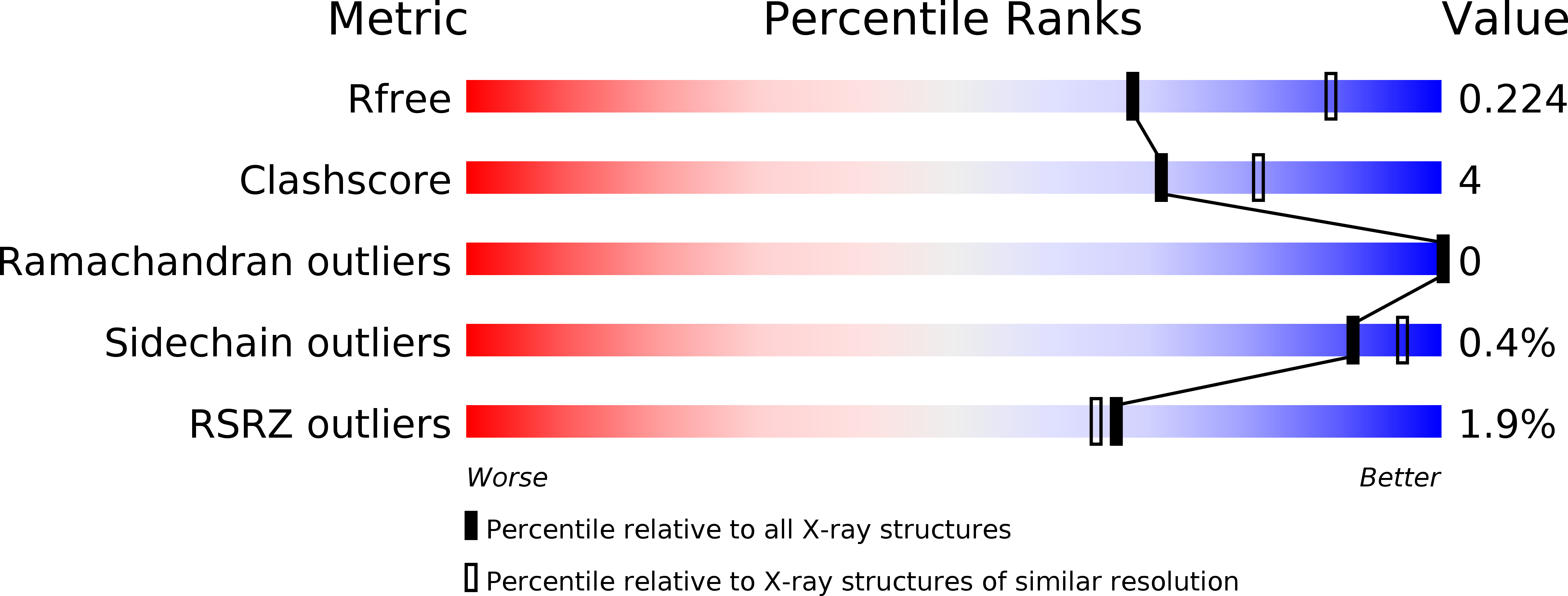
  • R-Value Free: 0.228 
  • R-Value Work: 0.173 
  • R-Value Observed: 0.176 

Starting Model: in silico
View more details

wwPDB Validation   3D Report Full Report


Ligand Structure Quality Assessment 


This is version 1.1 of the entry. See complete history


Literature

Crystal structure of the flavoenzyme PA4991 from Pseudomonas aeruginosa.

Jacewicz, A.Schnell, R.Lindqvist, Y.Schneider, G.

(2016) Acta Crystallogr F Struct Biol Commun 72: 105-111

  • DOI: https://doi.org/10.1107/S2053230X15024437
  • Primary Citation of Related Structures:  
    5EZ7

  • PubMed Abstract: 

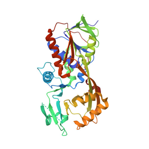
    The locus PA4991 in Pseudomonas aeruginosa encodes an open reading frame that has been identified as essential for the virulence and/or survival of this pathogenic organism in the infected host. Here, it is shown that this gene encodes a monomeric FAD-binding protein of molecular mass 42.2 kDa. The structure of PA4991 was determined by a combination of molecular replacement using a search model generated with Rosetta and phase improvement by a low-occupancy heavy-metal derivative. PA4991 belongs to the GR2 family of FAD-dependent oxidoreductases, comprising an FAD-binding domain typical of the glutathione reductase family and a second domain dominated by an eight-stranded mixed β-sheet. Most of the protein-FAD interactions are via the FAD-binding domain, but the isoalloxazine ring is located at the domain interface and interacts with residues from both domains. A comparison with the structurally related glycine oxidase and glycerol-3-phosphate dehydrogenase shows that in spite of very low amino-acid sequence identity (<18%) several active-site residues involved in substrate binding in these enzymes are conserved in PA4991. However, enzymatic assays show that PA4991 does not display amino-acid oxidase or glycerol-3-phosphate dehydrogenase activities, suggesting that it requires different substrates for activity.


  • Organizational Affiliation

    Department of Medical Biochemistry and Biophysics, Karolinska Institutet, S-171 77 Stockholm, Sweden.


Macromolecules
Find similar proteins by:  (by identity cutoff)  |  3D Structure
Entity ID: 1
MoleculeChains Sequence LengthOrganismDetailsImage
flavoenzyme PA4991392Pseudomonas aeruginosa PAO1Mutation(s): 0 
Gene Names: PA4991
EC: 1
UniProt
Find proteins for Q9HUH4 (Pseudomonas aeruginosa (strain ATCC 15692 / DSM 22644 / CIP 104116 / JCM 14847 / LMG 12228 / 1C / PRS 101 / PAO1))
Explore Q9HUH4 
Go to UniProtKB:  Q9HUH4
Entity Groups  
Sequence Clusters30% Identity50% Identity70% Identity90% Identity95% Identity100% Identity
UniProt GroupQ9HUH4
Sequence Annotations
Expand
  • Reference Sequence
Experimental Data & Validation

Experimental Data

  • Method: X-RAY DIFFRACTION
  • Resolution: 2.40 Å
  • R-Value Free: 0.228 
  • R-Value Work: 0.173 
  • R-Value Observed: 0.176 
  • Space Group: P 1 21 1
Unit Cell:
Length ( Å )Angle ( ˚ )
a = 37.89α = 90
b = 79.83β = 104.4
c = 63.23γ = 90
Software Package:
Software NamePurpose
REFMACrefinement
MOSFLMdata reduction
SCALAdata scaling
PHASERphasing

Structure Validation

View Full Validation Report


Ligand Structure Quality Assessment 


Entry History & Funding Information

Deposition Data


Funding OrganizationLocationGrant Number
European CommissionSweden--
Swedish Research CouncilSweden--

Revision History  (Full details and data files)

  • Version 1.0: 2016-02-17
    Type: Initial release
  • Version 1.1: 2024-05-01
    Changes: Data collection, Database references, Refinement description