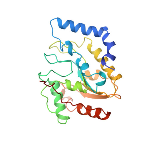
Crystal structure of Escherichia coli uracil DNA glycosylase and its complexes with uracil and glycerol: structure and glycosylase mechanism revisited.
Xiao, G., Tordova, M., Jagadeesh, J., Drohat, A.C., Stivers, J.T., Gilliland, G.L.(1999) Proteins 35: 13-24
- PubMed: 10090282
- Primary Citation of Related Structures:
1EUG, 2EUG, 3EUG, 5EUG - PubMed Abstract:
The DNA repair enzyme uracil DNA glycosylase (UDG) catalyzes the hydrolysis of premutagenic uracil residues from single-stranded or duplex DNA, producing free uracil and abasic DNA. Here we report the high-resolution crystal structures of free UDG from Escherichia coli strain B (1.60 A), its complex with uracil (1.50 A), and a second active-site complex with glycerol (1.43 A). These represent the first high-resolution structures of a prokaryotic UDG to be reported. The overall structure of the E. coli enzyme is more similar to the human UDG than the herpes virus enzyme. Significant differences between the bacterial and viral structures are seen in the side-chain positions of the putative general-acid (His187) and base (Asp64), similar to differences previously observed between the viral and human enzymes. In general, the active-site loop that contains His187 appears preorganized in comparison with the viral and human enzymes, requiring smaller substrate-induced conformational changes to bring active-site groups into catalytic position. These structural differences may be related to the large differences in the mechanism of uracil recognition used by the E. coli and viral enzymes. The pH dependence of k(cat) for wild-type UDG and the D64N and H187Q mutant enzymes is consistent with general-base catalysis by Asp64, but provides no evidence for a general-acid catalyst. The catalytic mechanism of UDG is critically discussed with respect to these results.
Organizational Affiliation:
Center for Advanced Research in Biotechnology, University of Maryland Biotechnology Institute and the National Institute for Standards and Technology, Rockville, 20850, USA.


