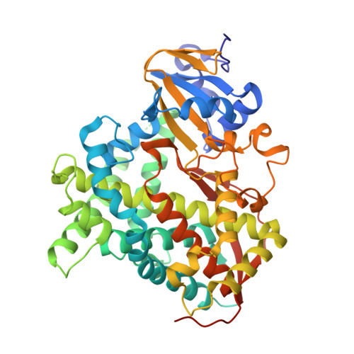
5DYZ

Crystal structure of Asp251Gly/Gln307His mutant of cytochrome P450 BM3 in complex with N-palmitoylglycine


Experimental Data Snapshot

  • Method: X-RAY DIFFRACTION
  • Resolution: 1.97 Å
  • R-Value Free: 0.219 
  • R-Value Work: 0.179 
  • R-Value Observed: 0.181 

Starting Model: experimental
View more details

wwPDB Validation   3D Report Full Report


Ligand Structure Quality Assessment 


This is version 1.2 of the entry. See complete history


Literature

Subtle structural changes in the Asp251Gly/Gln307His P450 BM3 mutant responsible for new activity toward diclofenac, tolbutamide and ibuprofen.

Di Nardo, G.Dell'Angelo, V.Catucci, G.Sadeghi, S.J.Gilardi, G.

(2016) Arch Biochem Biophys 602: 106-115

  • DOI: https://doi.org/10.1016/j.abb.2015.12.005
  • Primary Citation of Related Structures:  
    5DYP, 5DYZ

  • PubMed Abstract: 

    This paper reports the structure of the double mutant Asp251Gly/Gln307His (named A2) generated by random mutagenesis, able to produce 4'-hydroxydiclofenac, 2-hydroxyibuprofen and 4-hydroxytolbutamide from diclofenac, ibuprofen and tolbutamide, respectively. The 3D structure of the substrate-free mutant shows a conformation similar to the closed one found in the substrate-bound wild type enzyme, but with a higher degree of disorder in the region of the G-helix and F-G loop. This is due to the mutation Asp251Gly that breaks the salt bridge between Aps251 on I-helix and Lys224 on G-helix, allowing the G-helix to move away from I-helix and conferring a higher degree of flexibility to this element. This subtle structural change is accompanied by long-range structural rearrangements of the active site with the rotation of Phe87 and a reorganization of catalytically important water molecules. The impact of these structural features on thermal stability, reduction potential and electron transfer is investigated. The data demonstrate that a single mutation far from the active site triggers an increase in protein flexibility in a key region, shifting the conformational equilibrium toward the closed form that is ready to accept electrons and enter the P450 catalytic cycle as soon as a substrate is accepted.


  • Organizational Affiliation

    Department of Life Sciences and Systems Biology, University of Torino, Via Accademia Albertina 13, Torino, Italy; CrisDi, Interdepartmental Center for Crystallography, University of Torino, Via Pietro Giuria 7, Torino, Italy.


Macromolecules
Find similar proteins by:  (by identity cutoff)  |  3D Structure
Entity ID: 1
MoleculeChains Sequence LengthOrganismDetailsImage
Bifunctional P-450/NADPH-P450 reductaseA,
B [auth C]
470Priestia megateriumMutation(s): 2 
Gene Names: cyp102A1cyp102
EC: 1.14.14.1 (PDB Primary Data), 1.6.2.4 (PDB Primary Data)
UniProt
Find proteins for P14779 (Priestia megaterium (strain ATCC 14581 / DSM 32 / CCUG 1817 / JCM 2506 / NBRC 15308 / NCIMB 9376 / NCTC 10342 / NRRL B-14308 / VKM B-512 / Ford 19))
Explore P14779 
Go to UniProtKB:  P14779
Entity Groups  
Sequence Clusters30% Identity50% Identity70% Identity90% Identity95% Identity100% Identity
UniProt GroupP14779
Sequence Annotations
Expand
  • Reference Sequence
Small Molecules
Binding Affinity Annotations 
IDSourceBinding Affinity
140 BindingDB:  5DYZ Kd: min: 4, max: 297 (nM) from 4 assay(s)
Experimental Data & Validation

Experimental Data

  • Method: X-RAY DIFFRACTION
  • Resolution: 1.97 Å
  • R-Value Free: 0.219 
  • R-Value Work: 0.179 
  • R-Value Observed: 0.181 
  • Space Group: P 21 21 21
Unit Cell:
Length ( Å )Angle ( ˚ )
a = 61.213α = 90
b = 115.708β = 90
c = 143.278γ = 90
Software Package:
Software NamePurpose
PHENIXrefinement
XDSdata reduction
Aimlessdata scaling
MOLREPphasing

Structure Validation

View Full Validation Report


Ligand Structure Quality Assessment 


Entry History 

Deposition Data

Revision History  (Full details and data files)

  • Version 1.0: 2016-01-20
    Type: Initial release
  • Version 1.1: 2016-06-22
    Changes: Database references
  • Version 1.2: 2024-01-10
    Changes: Data collection, Database references, Refinement description