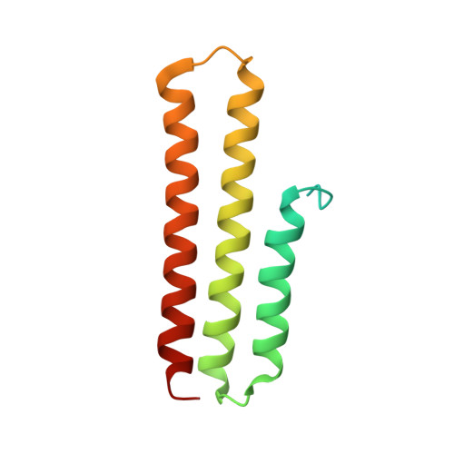
5DWK

Diacylglycerol Kinase solved by multi crystal multi orientation native SAD


Experimental Data Snapshot

  • Method: X-RAY DIFFRACTION
  • Resolution: 2.60 Å
  • R-Value Free: 0.245 
  • R-Value Work: 0.201 
  • R-Value Observed: 0.204 

wwPDB Validation   3D Report Full Report


Ligand Structure Quality Assessment 


This is version 1.1 of the entry. See complete history


Literature

Data-collection strategy for challenging native SAD phasing.

Olieric, V.Weinert, T.Finke, A.D.Anders, C.Li, D.Olieric, N.Borca, C.N.Steinmetz, M.O.Caffrey, M.Jinek, M.Wang, M.

(2016) Acta Crystallogr D Struct Biol 72: 421-429

  • DOI: https://doi.org/10.1107/S2059798315024110
  • Primary Citation of Related Structures:  
    5DWK, 5FQ5

  • PubMed Abstract: 

    Recent improvements in data-collection strategies have pushed the limits of native SAD (single-wavelength anomalous diffraction) phasing, a method that uses the weak anomalous signal of light elements naturally present in macromolecules. These involve the merging of multiple data sets from either multiple crystals or from a single crystal collected in multiple orientations at a low X-ray dose. Both approaches yield data of high multiplicity while minimizing radiation damage and systematic error, thus ensuring accurate measurements of the anomalous differences. Here, the combined use of these two strategies is described to solve cases of native SAD phasing that were particular challenges: the integral membrane diacylglycerol kinase (DgkA) with a low Bijvoet ratio of 1% and the large 200 kDa complex of the CRISPR-associated endonuclease (Cas9) bound to guide RNA and target DNA crystallized in the low-symmetry space group C2. The optimal native SAD data-collection strategy based on systematic measurements performed on the 266 kDa multiprotein/multiligand tubulin complex is discussed.


  • Organizational Affiliation

    Swiss Light Source, Paul Scherrer Institut, Villigen PSI, Switzerland.


Macromolecules
Find similar proteins by:  (by identity cutoff)  |  3D Structure
Entity ID: 1
MoleculeChains Sequence LengthOrganismDetailsImage
Diacylglycerol kinase
A, B, C, D, E
A, B, C, D, E, F
130Escherichia coli K-12Mutation(s): 7 
Gene Names: dgkAb4042JW4002
EC: 2.7.1.107
Membrane Entity: Yes 
UniProt
Find proteins for P0ABN1 (Escherichia coli (strain K12))
Explore P0ABN1 
Go to UniProtKB:  P0ABN1
Entity Groups  
Sequence Clusters30% Identity50% Identity70% Identity90% Identity95% Identity100% Identity
UniProt GroupP0ABN1
Sequence Annotations
Expand
  • Reference Sequence
Small Molecules
Ligands 6 Unique
IDChains Name / Formula / InChI Key2D Diagram3D Interactions
78M
Query on 78M

Download Ideal Coordinates CCD File 
J [auth A]
N [auth B]
P [auth C]
Q [auth C]
R [auth C]
J [auth A],
N [auth B],
P [auth C],
Q [auth C],
R [auth C],
S [auth C]
(2S)-2,3-DIHYDROXYPROPYL(7Z)-PENTADEC-7-ENOATE
C18 H34 O4
BJMLBVHMHXYQFS-JJEJIETFSA-N
78N
Query on 78N

Download Ideal Coordinates CCD File 
G [auth A]
H [auth A]
I [auth A]
K [auth B]
L [auth B]
G [auth A],
H [auth A],
I [auth A],
K [auth B],
L [auth B],
M [auth B],
W [auth D]
(2R)-2,3-DIHYDROXYPROPYL(7Z)-PENTADEC-7-ENOATE
C18 H34 O4
BJMLBVHMHXYQFS-XZVRFQMRSA-N
CIT
Query on CIT

Download Ideal Coordinates CCD File 
U [auth C]CITRIC ACID
C6 H8 O7
KRKNYBCHXYNGOX-UHFFFAOYSA-N
ZN
Query on ZN

Download Ideal Coordinates CCD File 
T [auth C]ZINC ION
Zn
PTFCDOFLOPIGGS-UHFFFAOYSA-N
ACT
Query on ACT

Download Ideal Coordinates CCD File 
V [auth C]ACETATE ION
C2 H3 O2
QTBSBXVTEAMEQO-UHFFFAOYSA-M
NA
Query on NA

Download Ideal Coordinates CCD File 
O [auth B]SODIUM ION
Na
FKNQFGJONOIPTF-UHFFFAOYSA-N
Experimental Data & Validation

Experimental Data

  • Method: X-RAY DIFFRACTION
  • Resolution: 2.60 Å
  • R-Value Free: 0.245 
  • R-Value Work: 0.201 
  • R-Value Observed: 0.204 
  • Space Group: P 21 21 21
Unit Cell:
Length ( Å )Angle ( ˚ )
a = 75.29α = 90
b = 91.57β = 90
c = 143.72γ = 90
Software Package:
Software NamePurpose
XDSdata reduction
XSCALEdata scaling
SHELXDEphasing
PHENIXrefinement

Structure Validation

View Full Validation Report


Ligand Structure Quality Assessment 


Entry History 

Deposition Data

Revision History  (Full details and data files)

  • Version 1.0: 2016-03-23
    Type: Initial release
  • Version 1.1: 2024-05-08
    Changes: Data collection, Database references