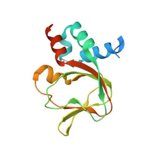
5C8W

PKG II's Amino Terminal Cyclic Nucleotide Binding Domain (CNB-A) in a complex with cGMP


Experimental Data Snapshot

  • Method: X-RAY DIFFRACTION
  • Resolution: 1.80 Å
  • R-Value Free: 0.196 
  • R-Value Work: 0.162 
  • R-Value Observed: 0.163 

wwPDB Validation   3D Report Full Report


Ligand Structure Quality Assessment 


This is version 1.2 of the entry. See complete history


Literature

Structural Basis of Cyclic Nucleotide Selectivity in cGMP-dependent Protein Kinase II.

Campbell, J.C.Kim, J.J.Li, K.Y.Huang, G.Y.Reger, A.S.Matsuda, S.Sankaran, B.Link, T.M.Yuasa, K.Ladbury, J.E.Casteel, D.E.Kim, C.

(2016) J Biol Chem 291: 5623-5633

  • DOI: https://doi.org/10.1074/jbc.M115.691303
  • Primary Citation of Related Structures:  
    5BV6, 5C6C, 5C8W

  • PubMed Abstract: 

    Membrane-bound cGMP-dependent protein kinase (PKG) II is a key regulator of bone growth, renin secretion, and memory formation. Despite its crucial physiological roles, little is known about its cyclic nucleotide selectivity mechanism due to a lack of structural information. Here, we find that the C-terminal cyclic nucleotide binding (CNB-B) domain of PKG II binds cGMP with higher affinity and selectivity when compared with its N-terminal CNB (CNB-A) domain. To understand the structural basis of cGMP selectivity, we solved co-crystal structures of the CNB domains with cyclic nucleotides. Our structures combined with mutagenesis demonstrate that the guanine-specific contacts at Asp-412 and Arg-415 of the αC-helix of CNB-B are crucial for cGMP selectivity and activation of PKG II. Structural comparison with the cGMP selective CNB domains of human PKG I and Plasmodium falciparum PKG (PfPKG) shows different contacts with the guanine moiety, revealing a unique cGMP selectivity mechanism for PKG II.


  • Organizational Affiliation

    From the Structural and Computational Biology and Molecular Biophysics Program.


Macromolecules
Find similar proteins by:  (by identity cutoff)  |  3D Structure
Entity ID: 1
MoleculeChains Sequence LengthOrganismDetailsImage
cGMP-dependent protein kinase 2
A, B, C, D, E
A, B, C, D, E, F
143Homo sapiensMutation(s): 0 
Gene Names: PRKG2PRKGR2
EC: 2.7.11.12
UniProt & NIH Common Fund Data Resources
Find proteins for Q13237 (Homo sapiens)
Explore Q13237 
Go to UniProtKB:  Q13237
PHAROS:  Q13237
GTEx:  ENSG00000138669 
Entity Groups  
Sequence Clusters30% Identity50% Identity70% Identity90% Identity95% Identity100% Identity
UniProt GroupQ13237
Sequence Annotations
Expand
  • Reference Sequence
Small Molecules
Ligands 3 Unique
IDChains Name / Formula / InChI Key2D Diagram3D Interactions
PCG
Query on PCG

Download Ideal Coordinates CCD File 
H [auth A]
L [auth B]
N [auth C]
Q [auth D]
S [auth E]
H [auth A],
L [auth B],
N [auth C],
Q [auth D],
S [auth E],
U [auth F]
CYCLIC GUANOSINE MONOPHOSPHATE
C10 H12 N5 O7 P
ZOOGRGPOEVQQDX-UUOKFMHZSA-N
MLA
Query on MLA

Download Ideal Coordinates CCD File 
G [auth A]
J [auth B]
K [auth B]
M [auth C]
P [auth D]
G [auth A],
J [auth B],
K [auth B],
M [auth C],
P [auth D],
R [auth E],
T [auth F]
MALONIC ACID
C3 H4 O4
OFOBLEOULBTSOW-UHFFFAOYSA-N
NA
Query on NA

Download Ideal Coordinates CCD File 
I [auth A],
O [auth C],
V [auth F]
SODIUM ION
Na
FKNQFGJONOIPTF-UHFFFAOYSA-N
Experimental Data & Validation

Experimental Data

  • Method: X-RAY DIFFRACTION
  • Resolution: 1.80 Å
  • R-Value Free: 0.196 
  • R-Value Work: 0.162 
  • R-Value Observed: 0.163 
  • Space Group: P 21 21 21
Unit Cell:
Length ( Å )Angle ( ˚ )
a = 46.71α = 90
b = 103.454β = 90
c = 176.405γ = 90
Software Package:
Software NamePurpose
PHENIXrefinement
HKL-3000data reduction
HKL-3000data scaling
PHASERphasing

Structure Validation

View Full Validation Report


Ligand Structure Quality Assessment 


Entry History 

Deposition Data

Revision History  (Full details and data files)

  • Version 1.0: 2016-01-20
    Type: Initial release
  • Version 1.1: 2016-04-27
    Changes: Database references
  • Version 1.2: 2024-03-06
    Changes: Data collection, Database references, Derived calculations