Towards phasing using high X-ray intensity.
Galli, L., Son, S.K., Barends, T.R., White, T.A., Barty, A., Botha, S., Boutet, S., Caleman, C., Doak, R.B., Nanao, M.H., Nass, K., Shoeman, R.L., Timneanu, N., Santra, R., Schlichting, I., Chapman, H.N.(2015) IUCrJ 2: 627-634
- PubMed: 26594370
- DOI: https://doi.org/10.1107/S2052252515014049
- Primary Citation of Related Structures:
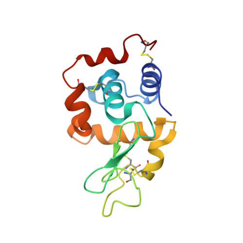
5C6I, 5C6J, 5C6L - PubMed Abstract:
X-ray free-electron lasers (XFELs) show great promise for macromolecular structure determination from sub-micrometre-sized crystals, using the emerging method of serial femtosecond crystallography. The extreme brightness of the XFEL radiation can multiply ionize most, if not all, atoms in a protein, causing their scattering factors to change during the pulse, with a preferential 'bleaching' of heavy atoms. This paper investigates the effects of electronic damage on experimental data collected from a Gd derivative of lysozyme microcrystals at different X-ray intensities, and the degree of ionization of Gd atoms is quantified from phased difference Fourier maps. A pattern sorting scheme is proposed to maximize the ionization contrast and the way in which the local electronic damage can be used for a new experimental phasing method is discussed.
Organizational Affiliation:
Center for Free-Electron Laser Science, Deutsches Elektronen-Synchrotron DESY , Notkestrasse 85, Hamburg, 22607, Germany ; Department of Physics, University of Hamburg , Luruper Chaussee 149, Hamburg, 22761, Germany.