5BY6

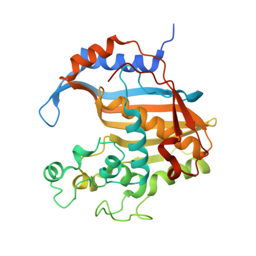
Crystal structure of Trichinella spiralis thymidylate synthase complexed with dUMP


Experimental Data Snapshot

  • Method: X-RAY DIFFRACTION
  • Resolution: 1.90 Å
  • R-Value Free: 0.220 
  • R-Value Work: 0.165 
  • R-Value Observed: 0.167 

Starting Model: experimental
View more details

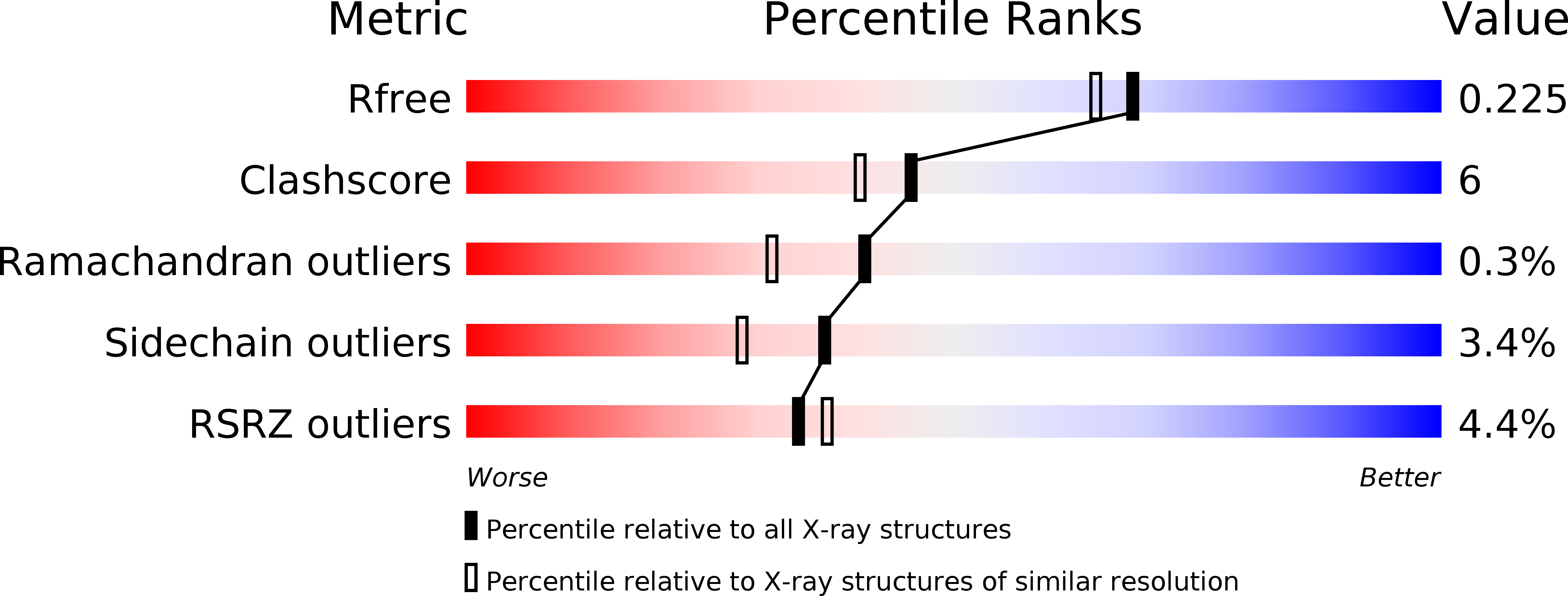
wwPDB Validation   3D Report Full Report


Ligand Structure Quality Assessment 


This is version 1.2 of the entry. See complete history


Literature

Crystal structures of nematode (parasitic T. spiralis and free living C. elegans), compared to mammalian, thymidylate synthases (TS). Molecular docking and molecular dynamics simulations in search for nematode-specific inhibitors of TS.

Jarmula, A.Wilk, P.Maj, P.Ludwiczak, J.Dowiercial, A.Banaszak, K.Rypniewski, W.Ciesla, J.Dabrowska, M.Fraczyk, T.Bronowska, A.K.Jakowiecki, J.Filipek, S.Rode, W.

(2017) J Mol Graph Model 77: 33-50

  • DOI: https://doi.org/10.1016/j.jmgm.2017.08.008
  • Primary Citation of Related Structures:  
    4IQB, 5BY6, 5NOO

  • PubMed Abstract: 

    Three crystal structures are presented of nematode thymidylate synthases (TS), including Caenorhabditis elegans (Ce) enzyme without ligands and its ternary complex with dUMP and Raltitrexed, and binary complex of Trichinella spiralis (Ts) enzyme with dUMP. In search of differences potentially relevant for the development of species-specific inhibitors of the nematode enzyme, a comparison was made of the present Ce and Ts enzyme structures, as well as binary complex of Ce enzyme with dUMP, with the corresponding mammalian (human, mouse and rat) enzyme crystal structures. To complement the comparison, tCONCOORD computations were performed to evaluate dynamic behaviors of mammalian and nematode TS structures. Finally, comparative molecular docking combined with molecular dynamics and free energy of binding calculations were carried out to search for ligands showing selective affinity to T. spiralis TS. Despite an overall strong similarity in structure and dynamics of nematode vs mammalian TSs, a pool of ligands demonstrating predictively a strong and selective binding to TsTS has been delimited. These compounds, the E63 family, locate in the dimerization interface of TsTS where they exert species-specific interactions with certain non-conserved residues, including hydrogen bonds with Thr174 and hydrophobic contacts with Phe192, Cys191 and Tyr152. The E63 family of ligands opens the possibility of future development of selective inhibitors of TsTS and effective agents against trichinellosis.


  • Organizational Affiliation

    Nencki Institute of Experimental Biology, Polish Academy of Sciences, Warszawa, Poland. Electronic address: a.jarmula@nencki.gov.pl.


Macromolecules
Find similar proteins by:  (by identity cutoff)  |  3D Structure
Entity ID: 1
MoleculeChains Sequence LengthOrganismDetailsImage
Thymidylate synthase
A, B, C, D
307Trichinella spiralisMutation(s): 0 
Gene Names: tsTsp_03568
EC: 2.1.1.45
UniProt
Find proteins for Q9NDD3 (Trichinella spiralis)
Explore Q9NDD3 
Go to UniProtKB:  Q9NDD3
Entity Groups  
Sequence Clusters30% Identity50% Identity70% Identity90% Identity95% Identity100% Identity
UniProt GroupQ9NDD3
Sequence Annotations
Expand
  • Reference Sequence
Small Molecules
Ligands 3 Unique
IDChains Name / Formula / InChI Key2D Diagram3D Interactions
UMP
Query on UMP

Download Ideal Coordinates CCD File 
E [auth A],
K [auth B],
L [auth C],
N [auth D]
2'-DEOXYURIDINE 5'-MONOPHOSPHATE
C9 H13 N2 O8 P
JSRLJPSBLDHEIO-SHYZEUOFSA-N
DTT
Query on DTT

Download Ideal Coordinates CCD File 
F [auth A]2,3-DIHYDROXY-1,4-DITHIOBUTANE
C4 H10 O2 S2
VHJLVAABSRFDPM-IMJSIDKUSA-N
GOL
Query on GOL

Download Ideal Coordinates CCD File 
G [auth A]
H [auth A]
I [auth A]
J [auth A]
M [auth C]
G [auth A],
H [auth A],
I [auth A],
J [auth A],
M [auth C],
O [auth D],
P [auth D],
Q [auth D]
GLYCEROL
C3 H8 O3
PEDCQBHIVMGVHV-UHFFFAOYSA-N
Experimental Data & Validation

Experimental Data

  • Method: X-RAY DIFFRACTION
  • Resolution: 1.90 Å
  • R-Value Free: 0.220 
  • R-Value Work: 0.165 
  • R-Value Observed: 0.167 
  • Space Group: P 1
Unit Cell:
Length ( Å )Angle ( ˚ )
a = 51.695α = 85.31
b = 65.914β = 85.33
c = 96.511γ = 67.12
Software Package:
Software NamePurpose
DENZOdata reduction
SCALEPACKdata scaling
PHASERphasing
REFMACrefinement
PDB_EXTRACTdata extraction
PHASERphasing

Structure Validation

View Full Validation Report


Ligand Structure Quality Assessment 


Entry History 

Deposition Data

Revision History  (Full details and data files)

  • Version 1.0: 2015-06-17
    Type: Initial release
  • Version 1.1: 2017-12-06
    Changes: Database references
  • Version 1.2: 2024-01-10
    Changes: Data collection, Database references, Refinement description