5YHT

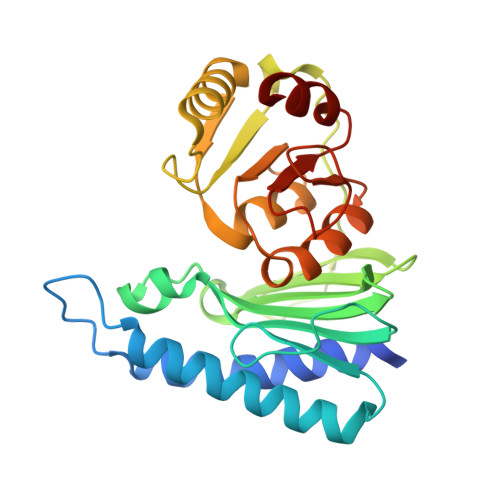
Crystal structure of a phosphatase from Mycobacterium tuberculosis in complex with its substrate


Experimental Data Snapshot

  • Method: X-RAY DIFFRACTION
  • Resolution: 2.87 Å
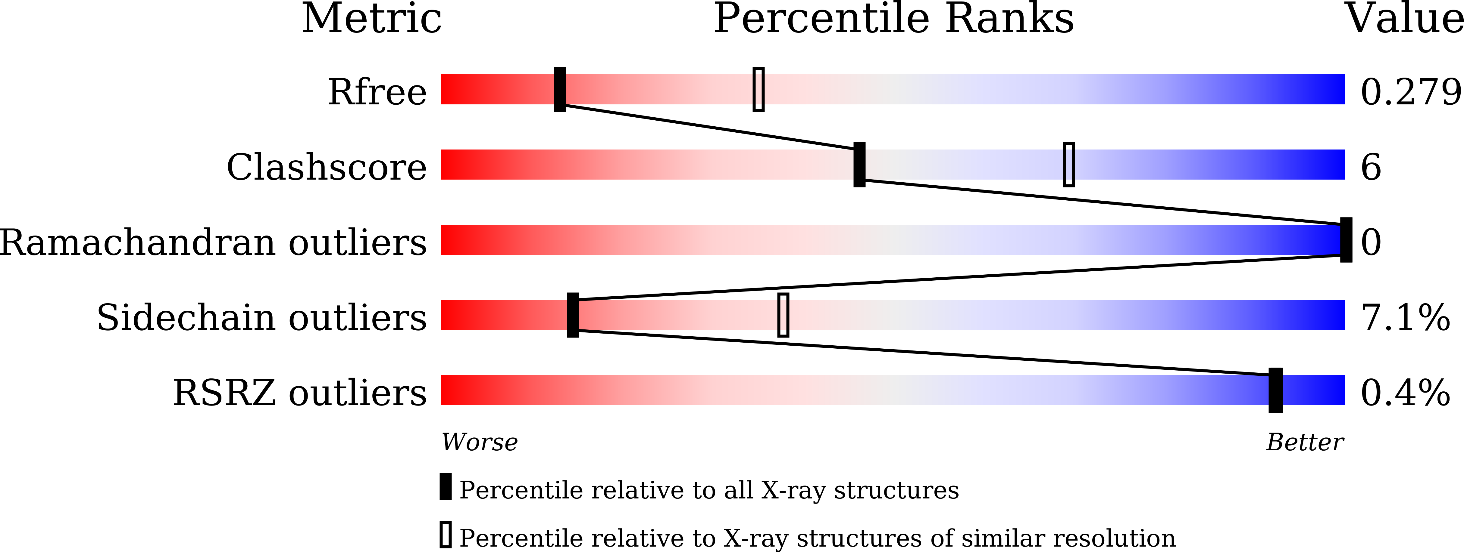
  • R-Value Free: 0.279 
  • R-Value Work: 0.229 
  • R-Value Observed: 0.233 

Starting Model: experimental
View more details

wwPDB Validation   3D Report Full Report


Ligand Structure Quality Assessment 


This is version 1.3 of the entry. See complete history


Literature

Identification and structural characterization of a histidinol phosphate phosphatase fromMycobacterium tuberculosis

Jha, B.Kumar, D.Sharma, A.Dwivedy, A.Singh, R.Biswal, B.K.

(2018) J Biol Chem 293: 10102-10118

  • DOI: https://doi.org/10.1074/jbc.RA118.002299
  • Primary Citation of Related Structures:  
    5YHT, 5ZON

  • PubMed Abstract: 

    The absence of a histidine biosynthesis pathway in humans, coupled with histidine essentiality for survival of the important human pathogen Mycobacterium tuberculosis ( Mtb ), underscores the importance of the bacterial enzymes of this pathway as major antituberculosis drug targets. However, the identity of the mycobacterial enzyme that functions as the histidinol phosphate phosphatase (HolPase) of this pathway remains to be established. Here, we demonstrate that the enzyme encoded by the Rv3137 gene, belonging to the inositol monophosphatase (IMPase) family, functions as the Mtb HolPase and specifically dephosphorylates histidinol phosphate. The crystal structure of Rv3137 in apo form enabled us to dissect its distinct structural features. Furthermore, the holo-complex structure revealed that a unique cocatalytic multizinc-assisted mode of substrate binding and catalysis is the hallmark of Mtb HolPase. Interestingly, the enzyme-substrate complex structure unveiled that although monomers possess individual catalytic sites they share a common product-exit channel at the dimer interface. Furthermore, target-based screening against HolPase identified several small-molecule inhibitors of this enzyme. Taken together, our study unravels the missing enzyme link in the Mtb histidine biosynthesis pathway, augments our current mechanistic understanding of histidine production in Mtb , and has helped identify potential inhibitors of this bacterial pathway.


  • Organizational Affiliation

    From the Structural and Functional Biology Laboratory, National Institute of Immunology, Aruna Asaf Ali Marg, New Delhi, Delhi 110067, India and.


Macromolecules
Find similar proteins by:  (by identity cutoff)  |  3D Structure
Entity ID: 1
MoleculeChains Sequence LengthOrganismDetailsImage
Histidinol-phosphatase
A, B
267Mycobacterium tuberculosis H37RvMutation(s): 0 
Gene Names: hisNimpCRv3137
EC: 3.1.3.15
UniProt
Find proteins for P95189 (Mycobacterium tuberculosis (strain ATCC 25618 / H37Rv))
Explore P95189 
Go to UniProtKB:  P95189
Entity Groups  
Sequence Clusters30% Identity50% Identity70% Identity90% Identity95% Identity100% Identity
UniProt GroupP95189
Sequence Annotations
Expand
  • Reference Sequence
Small Molecules
Ligands 4 Unique
IDChains Name / Formula / InChI Key2D Diagram3D Interactions
HSA
Query on HSA

Download Ideal Coordinates CCD File 
E [auth A]PHOSPHORIC ACID MONO-[2-AMINO-3-(3H-IMIDAZOL-4-YL)-PROPYL]ESTER
C6 H12 N3 O4 P
CWNDERHTHMWBSI-YFKPBYRVSA-N
HSO
Query on HSO

Download Ideal Coordinates CCD File 
G [auth B]L-histidinol
C6 H12 N3 O
ZQISRDCJNBUVMM-YFKPBYRVSA-O
PO4
Query on PO4

Download Ideal Coordinates CCD File 
F [auth A],
J [auth B],
K [auth B]
PHOSPHATE ION
O4 P
NBIIXXVUZAFLBC-UHFFFAOYSA-K
ZN
Query on ZN

Download Ideal Coordinates CCD File 
C [auth A],
D [auth A],
H [auth B],
I [auth B]
ZINC ION
Zn
PTFCDOFLOPIGGS-UHFFFAOYSA-N
Experimental Data & Validation

Experimental Data

  • Method: X-RAY DIFFRACTION
  • Resolution: 2.87 Å
  • R-Value Free: 0.279 
  • R-Value Work: 0.229 
  • R-Value Observed: 0.233 
  • Space Group: P 21 21 2
Unit Cell:
Length ( Å )Angle ( ˚ )
a = 92.664α = 90
b = 142.446β = 90
c = 48.723γ = 90
Software Package:
Software NamePurpose
REFMACrefinement
HKL-2000data reduction
HKL-2000data scaling
PHASERphasing

Structure Validation

View Full Validation Report


Ligand Structure Quality Assessment 


Entry History & Funding Information

Deposition Data


Funding OrganizationLocationGrant Number
Department of BiotechnologyIndia--

Revision History  (Full details and data files)

  • Version 1.0: 2018-05-23
    Type: Initial release
  • Version 1.1: 2018-05-30
    Changes: Data collection, Database references
  • Version 1.2: 2018-07-11
    Changes: Data collection, Database references, Source and taxonomy
  • Version 1.3: 2023-11-22
    Changes: Data collection, Database references, Derived calculations, Refinement description