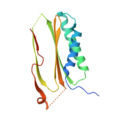
5WI4

CRYSTAL STRUCTURE OF DYNLT1/TCTEX-1 IN COMPLEX WITH ARHGEF2


Experimental Data Snapshot

  • Method: X-RAY DIFFRACTION
  • Resolution: 2.00 Å
  • R-Value Free: 0.210 
  • R-Value Work: 0.174 
  • R-Value Observed: 0.176 

Starting Model: experimental
View more details

wwPDB Validation   3D Report Full Report


This is version 1.1 of the entry. See complete history


Literature

MARK3-mediated phosphorylation of ARHGEF2 couples microtubules to the actin cytoskeleton to establish cell polarity.

Sandi, M.J.Marshall, C.B.Balan, M.Coyaud, E.Zhou, M.Monson, D.M.Ishiyama, N.Chandrakumar, A.A.La Rose, J.Couzens, A.L.Gingras, A.C.Raught, B.Xu, W.Ikura, M.Morrison, D.K.Rottapel, R.

(2017) Sci Signal 10

  • DOI: https://doi.org/10.1126/scisignal.aan3286
  • Primary Citation of Related Structures:  
    5WI4

  • PubMed Abstract: 

    The PAR-1-MARK pathway controls cell polarity through the phosphorylation of microtubule-associated proteins. Rho-Rac guanine nucleotide exchange factor 2 (ARHGEF2), which activates Ras homolog family member A (RHOA), is anchored to the microtubule network and sequestered in an inhibited state through binding to dynein light chain Tctex-1 type 1 (DYNLT1). We showed in mammalian cells that liver kinase B1 (LKB1) activated the microtubule affinity-regulating kinase 3 (MARK3), which in turn phosphorylated ARHGEF2 at Ser 151 This modification disrupted the interaction between ARHGEF2 and DYNLT1 by generating a 14-3-3 binding site in ARHGEF2, thus causing ARHGEF2 to dissociate from microtubules. Phosphorylation of ARHGEF2 by MARK3 stimulated RHOA activation and the formation of stress fibers and focal adhesions, and was required for organized cellular architecture in three-dimensional culture. Protein phosphatase 2A (PP2A) dephosphorylated Ser 151 in ARHGEF2 to restore the inhibited state. Thus, we have identified a regulatory switch controlled by MARK3 that couples microtubules to the actin cytoskeleton to establish epithelial cell polarity through ARHGEF2.


  • Organizational Affiliation

    Princess Margaret Cancer Centre, University Health Network, 101 College Street, Princess Margaret Cancer Research Tower, Toronto, Ontario M5G 1L7, Canada.


Macromolecules
Find similar proteins by:  (by identity cutoff)  |  3D Structure
Entity ID: 1
MoleculeChains Sequence LengthOrganismDetailsImage
Dynein light chain Tctex-type 1,Rho guanine nucleotide exchange factor 2
A, B, C
145Mus musculusMutation(s): 0 
Gene Names: Dynlt1Tctel1Tctex-1Tctex1Arhgef2Kiaa0651Lbcl1Lfc
UniProt & NIH Common Fund Data Resources
Find proteins for Q60875 (Mus musculus)
Explore Q60875 
Go to UniProtKB:  Q60875
IMPC:  MGI:103264
Find proteins for P51807 (Mus musculus)
Explore P51807 
Go to UniProtKB:  P51807
Entity Groups  
Sequence Clusters30% Identity50% Identity70% Identity90% Identity95% Identity100% Identity
UniProt GroupsP51807Q60875
Sequence Annotations
Expand
  • Reference Sequence
Experimental Data & Validation

Experimental Data

  • Method: X-RAY DIFFRACTION
  • Resolution: 2.00 Å
  • R-Value Free: 0.210 
  • R-Value Work: 0.174 
  • R-Value Observed: 0.176 
  • Space Group: P 32 2 1
Unit Cell:
Length ( Å )Angle ( ˚ )
a = 91.967α = 90
b = 91.967β = 90
c = 83.079γ = 120
Software Package:
Software NamePurpose
HKL-2000data reduction
HKL-2000data scaling
Cootphasing
PHENIXrefinement

Structure Validation

View Full Validation Report


Entry History & Funding Information

Deposition Data


Funding OrganizationLocationGrant Number
Cancer Research Society (CRS)CanadaCRS 14014
Canada Foundation for InnovationCanada--

Revision History  (Full details and data files)

  • Version 1.0: 2017-11-15
    Type: Initial release
  • Version 1.1: 2023-10-04
    Changes: Advisory, Data collection, Database references, Refinement description