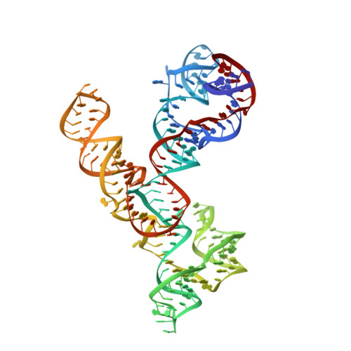
5V3I

Crystal structure of the VS ribozyme - wild-type C634

  • Classification: RNA
  • Organism(s): Neurospora
  • Mutation(s): No 

  • Deposited: 2017-03-07 Released: 2017-07-05 
  • Deposition Author(s): DasGupta, S., Suslov, N.B., Piccirilli, J.A.
  • Funding Organization(s): National Institutes of Health/National Institute Of Allergy and Infectious Diseases (NIH/NIAID)

Experimental Data Snapshot

  • Method: X-RAY DIFFRACTION
  • Resolution: 3.29 Å
  • R-Value Free: 0.246 
  • R-Value Work: 0.216 
  • R-Value Observed: 0.219 

Starting Model: experimental
View more details

wwPDB Validation   3D Report Full Report


This is version 1.3 of the entry. See complete history


Literature

Structural Basis for Substrate Helix Remodeling and Cleavage Loop Activation in the Varkud Satellite Ribozyme.

DasGupta, S.Suslov, N.B.Piccirilli, J.A.

(2017) J Am Chem Soc 139: 9591-9597

  • DOI: https://doi.org/10.1021/jacs.7b03655
  • Primary Citation of Related Structures:  
    5V3I

  • PubMed Abstract: 

    The Varkud satellite (VS) ribozyme catalyzes site-specific RNA cleavage and ligation reactions. Recognition of the substrate involves a kissing loop interaction between the substrate and the catalytic domain of the ribozyme, resulting in a rearrangement of the substrate helix register into a so-called "shifted" conformation that is critical for substrate binding and activation. We report a 3.3 Å crystal structure of the complete ribozyme that reveals the active, shifted conformation of the substrate, docked into the catalytic domain of the ribozyme. Comparison to previous NMR structures of isolated, inactive substrates provides a physical description of substrate remodeling, and implicates roles for tertiary interactions in catalytic activation of the cleavage loop. Similarities to the hairpin ribozyme cleavage loop activation suggest general strategies to enhance fidelity in RNA folding and ribozyme cleavage.


  • Organizational Affiliation

    Department of Chemistry, and ‡Department of Biochemistry and Molecular Biology, The University of Chicago , Chicago, Illinois 60637, United States.


Macromolecules
Find similar nucleic acids by:  (by identity cutoff)  |  3D Structure
Entity ID: 1
MoleculeChains LengthOrganismImage
VS Ribozyme RNA186Neurospora
Sequence Annotations
Expand
  • Reference Sequence
Experimental Data & Validation

Experimental Data

  • Method: X-RAY DIFFRACTION
  • Resolution: 3.29 Å
  • R-Value Free: 0.246 
  • R-Value Work: 0.216 
  • R-Value Observed: 0.219 
  • Space Group: P 43 21 2
Unit Cell:
Length ( Å )Angle ( ˚ )
a = 104.306α = 90
b = 104.306β = 90
c = 211.906γ = 90
Software Package:
Software NamePurpose
PHENIXrefinement
XDSdata reduction
Aimlessdata scaling
PHENIXphasing

Structure Validation

View Full Validation Report


Entry History & Funding Information

Deposition Data


Funding OrganizationLocationGrant Number
National Institutes of Health/National Institute Of Allergy and Infectious Diseases (NIH/NIAID)United StatesRO1AI081987

Revision History  (Full details and data files)

  • Version 1.0: 2017-07-05
    Type: Initial release
  • Version 1.1: 2017-08-02
    Changes: Author supporting evidence, Database references
  • Version 1.2: 2019-12-11
    Changes: Author supporting evidence
  • Version 1.3: 2023-10-04
    Changes: Data collection, Database references, Refinement description