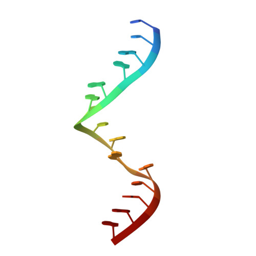
Nuclear Magnetic Resonance Structure of an 8 8 Nucleotide RNA Internal Loop Flanked on Each Side by Three Watson-Crick Pairs and Comparison to Three-Dimensional Predictions.
Kauffmann, A.D., Kennedy, S.D., Zhao, J., Turner, D.H.(2017) Biochemistry 56: 3733-3744
- PubMed: 28700212
- DOI: https://doi.org/10.1021/acs.biochem.7b00201
- Primary Citation of Related Structures:
5V2R - PubMed Abstract:
The prediction of RNA three-dimensional structure from sequence alone has been a long-standing goal. High-resolution, experimentally determined structures of simple noncanonical pairings and motifs are critical to the development of prediction programs. Here, we present the nuclear magnetic resonance structure of the (5'CCAGAAACGGAUGGA) 2 duplex, which contains an 8 × 8 nucleotide internal loop flanked by three Watson-Crick pairs on each side. The loop is comprised of a central 5'AC/3'CA nearest neighbor flanked by two 3RRs motifs, a known stable motif consisting of three consecutive sheared GA pairs. Hydrogen bonding patterns between base pairs in the loop, the all-atom root-mean-square deviation for the loop, and the deformation index were used to compare the structure to automated predictions by MC-sym, RNA FARFAR, and RNAComposer.
Organizational Affiliation:
Department of Chemistry, University of Rochester , Rochester, New York 14627, United States.


