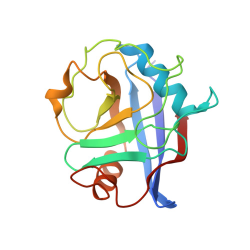
5T9Z

Discovery of a Potent Cyclophilin Inhibitor (Compound 6) based on Structural Simplification of Sanglifehrin A


Experimental Data Snapshot

  • Method: X-RAY DIFFRACTION
  • Resolution: 1.40 Å
  • R-Value Free: 0.190 
  • R-Value Work: 0.163 
  • R-Value Observed: 0.165 

wwPDB Validation   3D Report Full Report


Ligand Structure Quality Assessment 


This is version 1.4 of the entry. See complete history


Literature

Discovery of Potent Cyclophilin Inhibitors Based on the Structural Simplification of Sanglifehrin A.

Steadman, V.A.Pettit, S.B.Poullennec, K.G.Lazarides, L.Keats, A.J.Dean, D.K.Stanway, S.J.Austin, C.A.Sanvoisin, J.A.Watt, G.M.Fliri, H.G.Liclican, A.C.Jin, D.Wong, M.H.Leavitt, S.A.Lee, Y.J.Tian, Y.Frey, C.R.Appleby, T.C.Schmitz, U.Jansa, P.Mackman, R.L.Schultz, B.E.

(2017) J Med Chem 60: 1000-1017

  • DOI: https://doi.org/10.1021/acs.jmedchem.6b01329
  • Primary Citation of Related Structures:  
    5T9U, 5T9W, 5T9Z, 5TA2, 5TA4

  • PubMed Abstract: 

    Cyclophilin inhibition has been a target for the treatment of hepatitis C and other diseases, but the generation of potent, drug-like molecules through chemical synthesis has been challenging. In this study, a set of macrocyclic cyclophilin inhibitors was synthesized based on the core structure of the natural product sanglifehrin A. Initial compound optimization identified the valine-m-tyrosine-piperazic acid tripeptide (Val-m-Tyr-Pip) in the sanglifehrin core, stereocenters at C14 and C15, and the hydroxyl group of the m-tyrosine (m-Tyr) residue as key contributors to compound potency. Replacing the C18-C21 diene unit of sanglifehrin with a styryl group led to potent compounds that displayed a novel binding mode in which the styrene moiety engaged in a π-stacking interaction with Arg55 of cyclophilin A (Cyp A), and the m-Tyr residue was displaced into solvent. This observation allowed further simplifications of the scaffold to generate new lead compounds in the search for orally bioavailable cyclophilin inhibitors.


  • Organizational Affiliation

    Selcia Ltd. , Fyfield Business & Research Park, Fyfield Road, Ongar, Essex CM5 0GS, United Kingdom.


Macromolecules
Find similar proteins by:  (by identity cutoff)  |  3D Structure
Entity ID: 1
MoleculeChains Sequence LengthOrganismDetailsImage
Peptidyl-prolyl cis-trans isomerase A163Homo sapiensMutation(s): 0 
Gene Names: PPIACYPA
EC: 5.2.1.8
UniProt & NIH Common Fund Data Resources
Find proteins for P62937 (Homo sapiens)
Explore P62937 
Go to UniProtKB:  P62937
PHAROS:  P62937
GTEx:  ENSG00000196262 
Entity Groups  
Sequence Clusters30% Identity50% Identity70% Identity90% Identity95% Identity100% Identity
UniProt GroupP62937
Sequence Annotations
Expand
  • Reference Sequence
Small Molecules
Ligands 1 Unique
IDChains Name / Formula / InChI Key2D Diagram3D Interactions
78R
Query on 78R

Download Ideal Coordinates CCD File 
B [auth A]11-[(3-hydroxyphenyl)methyl]-18-methoxy-17-methyl-14-(propan-2-yl)-3-oxa-9,12,15,28-tetraazatricyclo[21.3.1.1~5,9~]octacosa-1(27),21,23,25-tetraene-4,10,13,16-tetrone
C35 H46 N4 O7
FLZFOCXCNGEIHF-HEPDUKFXSA-N
Experimental Data & Validation

Experimental Data

  • Method: X-RAY DIFFRACTION
  • Resolution: 1.40 Å
  • R-Value Free: 0.190 
  • R-Value Work: 0.163 
  • R-Value Observed: 0.165 
  • Space Group: P 21 21 21
Unit Cell:
Length ( Å )Angle ( ˚ )
a = 36.243α = 90
b = 51.865β = 90
c = 81.905γ = 90
Software Package:
Software NamePurpose
HKL-2000data collection
SCALEPACKdata scaling
PHENIXrefinement
PDB_EXTRACTdata extraction

Structure Validation

View Full Validation Report


Ligand Structure Quality Assessment 


Entry History 

Deposition Data

Revision History  (Full details and data files)

  • Version 1.0: 2017-01-25
    Type: Initial release
  • Version 1.1: 2017-02-08
    Changes: Database references
  • Version 1.2: 2017-02-15
    Changes: Database references
  • Version 1.3: 2017-03-29
    Changes: Structure summary
  • Version 1.4: 2024-03-06
    Changes: Data collection, Database references