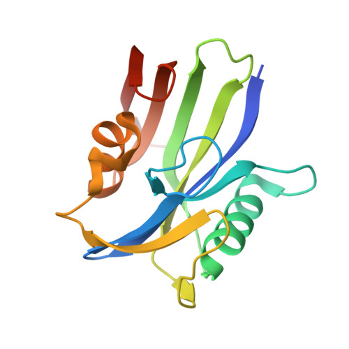
5NHY

BAY-707 in complex with MTH1


Experimental Data Snapshot

  • Method: X-RAY DIFFRACTION
  • Resolution: 1.72 Å
  • R-Value Free: 0.262 
  • R-Value Work: 0.220 
  • R-Value Observed: 0.222 

Starting Model: experimental
View more details

wwPDB Validation   3D Report Full Report


Ligand Structure Quality Assessment 


This is version 1.2 of the entry. See complete history


Literature

Novel Class of Potent and Cellularly Active Inhibitors Devalidates MTH1 as Broad-Spectrum Cancer Target.

Ellermann, M.Eheim, A.Rahm, F.Viklund, J.Guenther, J.Andersson, M.Ericsson, U.Forsblom, R.Ginman, T.Lindstrom, J.Silvander, C.Tresaugues, L.Giese, A.Bunse, S.Neuhaus, R.Weiske, J.Quanz, M.Glasauer, A.Nowak-Reppel, K.Bader, B.Irlbacher, H.Meyer, H.Queisser, N.Bauser, M.Haegebarth, A.Gorjanacz, M.

(2017) ACS Chem Biol 12: 1986-1992

  • DOI: https://doi.org/10.1021/acschembio.7b00370
  • Primary Citation of Related Structures:  
    5NHY

  • PubMed Abstract: 

    MTH1 is a hydrolase responsible for sanitization of oxidized purine nucleoside triphosphates to prevent their incorporation into replicating DNA. Early tool compounds published in the literature inhibited the enzymatic activity of MTH1 and subsequently induced cancer cell death; however recent studies have questioned the reported link between these two events. Therefore, it is important to validate MTH1 as a cancer dependency with high quality chemical probes. Here, we present BAY-707, a substrate-competitive, highly potent and selective inhibitor of MTH1, chemically distinct compared to those previously published. Despite superior cellular target engagement and pharmacokinetic properties, inhibition of MTH1 with BAY-707 resulted in a clear lack of in vitro or in vivo anticancer efficacy either in mono- or in combination therapies. Therefore, we conclude that MTH1 is dispensable for cancer cell survival.


  • Organizational Affiliation

    Bayer AG , Berlin, Germany.


Macromolecules
Find similar proteins by:  (by identity cutoff)  |  3D Structure
Entity ID: 1
MoleculeChains Sequence LengthOrganismDetailsImage
7,8-dihydro-8-oxoguanine triphosphatase
A, B
159Homo sapiensMutation(s): 0 
Gene Names: NUDT1MTH1
EC: 3.6.1.55 (PDB Primary Data), 3.6.1.56 (PDB Primary Data)
UniProt & NIH Common Fund Data Resources
Find proteins for P36639 (Homo sapiens)
Go to UniProtKB:  P36639
PHAROS:  P36639
GTEx:  ENSG00000106268 
Sequence Annotations
Expand
  • Reference Sequence
Small Molecules
Ligands 3 Unique
IDChains Name / Formula / InChI Key2D Diagram3D Interactions
8XT
Query on 8XT

Download Ideal Coordinates CCD File 
C [auth A],
K [auth B]
~{N}-ethyl-4-[(3~{S})-3-methylmorpholin-4-yl]-1~{H}-pyrrolo[2,3-b]pyridine-2-carboxamide
C15 H20 N4 O2
RPMGXDCRCWWCRY-JTQLQIEISA-N
SO4
Query on SO4

Download Ideal Coordinates CCD File 
D [auth A]
E [auth A]
F [auth A]
G [auth A]
H [auth A]
D [auth A],
E [auth A],
F [auth A],
G [auth A],
H [auth A],
I [auth A],
L [auth B]
SULFATE ION
O4 S
QAOWNCQODCNURD-UHFFFAOYSA-L
GOL
Query on GOL

Download Ideal Coordinates CCD File 
J [auth A],
M [auth B],
N [auth B]
GLYCEROL
C3 H8 O3
PEDCQBHIVMGVHV-UHFFFAOYSA-N
Binding Affinity Annotations 
IDSourceBinding Affinity
8XT BindingDB:  5NHY IC50: min: 0.8, max: 2.3 (nM) from 2 assay(s)
Experimental Data & Validation

Experimental Data

  • Method: X-RAY DIFFRACTION
  • Resolution: 1.72 Å
  • R-Value Free: 0.262 
  • R-Value Work: 0.220 
  • R-Value Observed: 0.222 
  • Space Group: P 21 21 21
Unit Cell:
Length ( Å )Angle ( ˚ )
a = 59.528α = 90
b = 66.709β = 90
c = 82.075γ = 90
Software Package:
Software NamePurpose
REFMACrefinement
XDSdata reduction
XDSdata scaling
PHASERphasing

Structure Validation

View Full Validation Report


Ligand Structure Quality Assessment 


Entry History 

Revision History  (Full details and data files)

  • Version 1.0: 2017-07-19
    Type: Initial release
  • Version 1.1: 2017-08-30
    Changes: Database references
  • Version 1.2: 2024-01-17
    Changes: Data collection, Database references, Refinement description