5N47

Structure of Anticalin N7E in complex with the three-domain fragment Fn7B8 of human oncofetal fibronectin


Experimental Data Snapshot

  • Method: X-RAY DIFFRACTION
  • Resolution: 3.00 Å
  • R-Value Free: 0.241 
  • R-Value Work: 0.208 
  • R-Value Observed: 0.210 

Starting Model: experimental
View more details

wwPDB Validation   3D Report Full Report


This is version 1.3 of the entry. See complete history


Literature

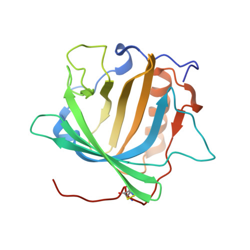
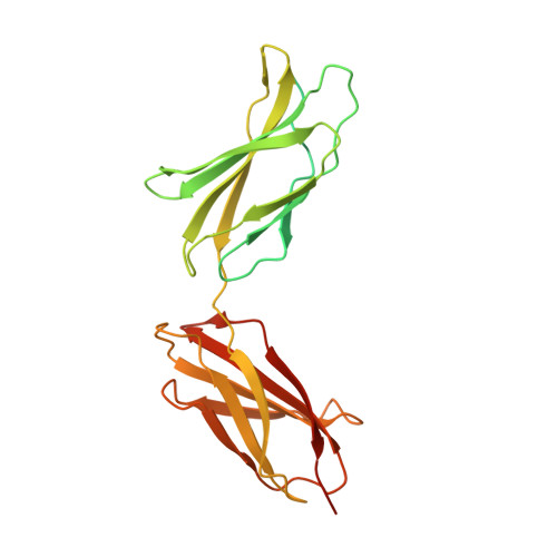
Anticalins Reveal High Plasticity in the Mode of Complex Formation with a Common Tumor Antigen.

Schiefner, A.Gebauer, M.Richter, A.Skerra, A.

(2018) Structure 26: 649-656.e3

  • DOI: https://doi.org/10.1016/j.str.2018.02.003
  • Primary Citation of Related Structures:  
    5N47, 5N48

  • PubMed Abstract: 

    We describe the comparative X-ray structural analysis of three Anticalin proteins directed against the extra-domain B (ED-B) of oncofetal fibronectin (Fn), a validated marker of tumor neoangiogenesis. The Anticalins were engineered from the human lipocalin 2 (Lcn2) scaffold via targeted randomization of the structurally variable loop region and selection by phage display, resulting in 15-19 exchanged residues. While the four reshaped loops exhibit diverse conformations (with shifts in Cα positions up to 20.4 Å), the β-barrel core of the lipocalin remains strongly conserved, thus confirming the extraordinary robustness of this scaffold. All three Anticalins bind the cc' hairpin loop of ED-B, the most exposed motif in the context of its neighboring Fn domains, but reveal entirely different binding modes, with orientations differing by up to 180°. Hence, each Anticalin recognizes its molecular target in an individual manner, in line with the distinct epitope specificities previously seen in binding experiments.


  • Organizational Affiliation

    Munich Center for Integrated Protein Science (CIPS-M) and Lehrstuhl für Biologische Chemie, Technische Universität München, Emil-Erlenmeyer-Forum 5, 85354 Freising (Weihenstephan), Germany.


Macromolecules
Find similar proteins by:  (by identity cutoff)  |  3D Structure
Entity ID: 1
MoleculeChains Sequence LengthOrganismDetailsImage
Neutrophil gelatinase-associated lipocalin
A, C, E
188Homo sapiensMutation(s): 17 
Gene Names: LCN2HNLNGAL
UniProt & NIH Common Fund Data Resources
Find proteins for P80188 (Homo sapiens)
Explore P80188 
Go to UniProtKB:  P80188
GTEx:  ENSG00000148346 
Entity Groups  
Sequence Clusters30% Identity50% Identity70% Identity90% Identity95% Identity100% Identity
UniProt GroupP80188
Sequence Annotations
Expand
  • Reference Sequence
Find similar proteins by:  (by identity cutoff)  |  3D Structure
Entity ID: 2
MoleculeChains Sequence LengthOrganismDetailsImage
Fibronectin
B, D, F
284Homo sapiensMutation(s): 0 
Gene Names: FN1FN
UniProt & NIH Common Fund Data Resources
Find proteins for P02751 (Homo sapiens)
Explore P02751 
Go to UniProtKB:  P02751
GTEx:  ENSG00000115414 
Entity Groups  
Sequence Clusters30% Identity50% Identity70% Identity90% Identity95% Identity100% Identity
UniProt GroupP02751
Sequence Annotations
Expand
  • Reference Sequence
Experimental Data & Validation

Experimental Data

  • Method: X-RAY DIFFRACTION
  • Resolution: 3.00 Å
  • R-Value Free: 0.241 
  • R-Value Work: 0.208 
  • R-Value Observed: 0.210 
  • Space Group: P 41 21 2
Unit Cell:
Length ( Å )Angle ( ˚ )
a = 135.148α = 90
b = 135.148β = 90
c = 203.939γ = 90
Software Package:
Software NamePurpose
XSCALEdata scaling
REFMACrefinement
PDB_EXTRACTdata extraction
XDSdata reduction
PHASERphasing

Structure Validation

View Full Validation Report


Entry History 

Deposition Data

Revision History  (Full details and data files)

  • Version 1.0: 2018-02-14
    Type: Initial release
  • Version 1.1: 2018-03-21
    Changes: Database references
  • Version 1.2: 2018-04-11
    Changes: Data collection, Database references
  • Version 1.3: 2024-01-17
    Changes: Data collection, Database references, Refinement description