5N0F

The catalytic domain, BcGH76, of Bacillus circulans Aman6 in complex with 1,6-ManSIFG


Experimental Data Snapshot

  • Method: X-RAY DIFFRACTION
  • Resolution: 1.69 Å
  • R-Value Free: 0.218 
  • R-Value Work: 0.152 
  • R-Value Observed: 0.156 

Starting Model: experimental
View more details

wwPDB Validation   3D Report Full Report


Ligand Structure Quality Assessment 


This is version 2.1 of the entry. See complete history


Literature

An atypical interaction explains the high-affinity of a non-hydrolyzable S-linked 1,6-alpha-mannanase inhibitor.

Belz, T.Jin, Y.Coines, J.Rovira, C.Davies, G.J.Williams, S.J.

(2017) Chem Commun (Camb) 53: 9238-9241

  • DOI: https://doi.org/10.1039/c7cc04977c
  • Primary Citation of Related Structures:  
    5M77, 5N0F

  • PubMed Abstract: 

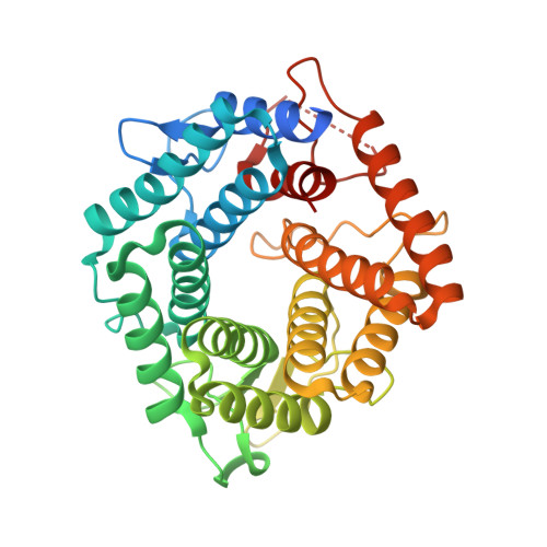
    The non-hydrolyzable S-linked azasugars, 1,6-α-mannosylthio- and 1,6-α-mannobiosylthioisofagomine, were synthesized and shown to bind with high affinity to a family 76 endo-1,6-α-mannanase from Bacillus circulans. X-ray crystallography showed an atypical interaction of the isofagomine nitrogen with the catalytic acid/base. Molecular dynamics simulations reveal that the atypical binding results from sulfur perturbing the most stable form away from the nucleophile interaction preferred for the O-linked congener.


  • Organizational Affiliation

    School of Chemistry and Bio21 Molecular Science and Biotechnology Institute, University of Melbourne, Parkville, Victoria 3010, Australia. sjwill@unimelb.edu.au.


Macromolecules
Find similar proteins by:  (by identity cutoff)  |  3D Structure
Entity ID: 1
MoleculeChains Sequence LengthOrganismDetailsImage
Alpha-1,6-mannanase
A, B
362Niallia circulansMutation(s): 1 
Gene Names: aman6
UniProt
Find proteins for Q9Z4P9 (Niallia circulans)
Explore Q9Z4P9 
Go to UniProtKB:  Q9Z4P9
Entity Groups  
Sequence Clusters30% Identity50% Identity70% Identity90% Identity95% Identity100% Identity
UniProt GroupQ9Z4P9
Sequence Annotations
Expand
  • Reference Sequence
Small Molecules
Ligands 1 Unique
IDChains Name / Formula / InChI Key2D Diagram3D Interactions
7K2
Query on 7K2

Download Ideal Coordinates CCD File 
C [auth A],
D [auth B]
[(3S,4R,5R)-4,5-dihydroxypiperidin-3-yl]methyl 1-thio-alpha-D-mannopyranoside
C12 H23 N O7 S
KDPQETHZRPVZAI-TYMLGRTHSA-N
Experimental Data & Validation

Experimental Data

  • Method: X-RAY DIFFRACTION
  • Resolution: 1.69 Å
  • R-Value Free: 0.218 
  • R-Value Work: 0.152 
  • R-Value Observed: 0.156 
  • Space Group: P 21 21 21
Unit Cell:
Length ( Å )Angle ( ˚ )
a = 85.05α = 90
b = 86.4β = 90
c = 103.65γ = 90
Software Package:
Software NamePurpose
REFMACrefinement
Aimlessdata scaling
XDSdata scaling
MOLREPphasing

Structure Validation

View Full Validation Report


Ligand Structure Quality Assessment 


Entry History & Funding Information

Deposition Data


Funding OrganizationLocationGrant Number
European Research CouncilUnited KingdomAdG322942
Australian Research CouncilAustraliaFT130100103

Revision History  (Full details and data files)

  • Version 1.0: 2017-08-09
    Type: Initial release
  • Version 1.1: 2017-08-16
    Changes: Database references
  • Version 1.2: 2017-08-23
    Changes: Database references
  • Version 2.0: 2020-07-29
    Type: Remediation
    Reason: Carbohydrate remediation
    Changes: Atomic model, Author supporting evidence, Derived calculations, Structure summary
  • Version 2.1: 2024-01-17
    Changes: Data collection, Database references, Refinement description, Structure summary