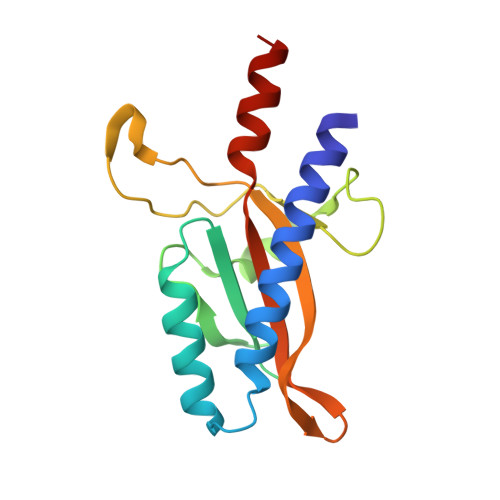
5KU5

Crystal Structure of CusS Sensor Domain with Silver Bound


Experimental Data Snapshot

  • Method: X-RAY DIFFRACTION
  • Resolution: 2.15 Å
  • R-Value Free: 0.286 
  • R-Value Work: 0.250 
  • R-Value Observed: 0.252 

wwPDB Validation   3D Report Full Report


This is version 1.4 of the entry. See complete history


Literature

The Structure of the Periplasmic Sensor Domain of the Histidine Kinase CusS Shows Unusual Metal Ion Coordination at the Dimeric Interface.

Affandi, T.Issaian, A.V.McEvoy, M.M.

(2016) Biochemistry 55: 5296-5306

  • DOI: https://doi.org/10.1021/acs.biochem.6b00707
  • Primary Citation of Related Structures:  
    5KU5

  • PubMed Abstract: 

    In bacteria, two-component systems act as signaling systems to respond to environmental stimuli. Two-component systems generally consist of a sensor histidine kinase and a response regulator, which work together through histidyl-aspartyl phosphorelay to result in gene regulation. One of the two-component systems in Escherichia coli, CusS-CusR, is known to induce expression of cusCFBA genes at increased periplasmic Cu(I) and Ag(I) concentrations to help maintain metal ion homeostasis. CusS is a membrane-associated histidine kinase with a periplasmic sensor domain connected to the cytoplasmic ATP binding and catalytic domains through two transmembrane helices. The mechanism of how CusS senses increasing metal ion concentrations and activates CusR is not yet known. Here, we present the crystal structure of the Ag(I)-bound periplasmic sensor domain of CusS at a resolution of 2.15 Å. The structure reveals that CusS forms a homodimer with four Ag(I) binding sites per dimeric complex. Two symmetric metal binding sites are found at the dimeric interface, which are each formed by two histidines and one phenylalanine with an unusual cation-π interaction. The other metal ion binding sites are in a nonconserved region within each monomer. Functional analyses of CusS variants with mutations in the metal sites suggest that the metal ion binding site at the dimer interface is more important for function. The structural and functional data provide support for a model in which metal-induced dimerization results in increases in kinase activity in the cytoplasmic domains of CusS.


  • Organizational Affiliation

    Department of Chemistry and Biochemistry, University of Arizona , Tucson, Arizona 85721, United States.


Macromolecules
Find similar proteins by:  (by identity cutoff)  |  3D Structure
Entity ID: 1
MoleculeChains Sequence LengthOrganismDetailsImage
Sensor kinase CusS
A, B, C, D
150Escherichia coli K-12Mutation(s): 0 
Gene Names: cusSybcZb0570JW5082
EC: 2.7.13.3
UniProt
Find proteins for P77485 (Escherichia coli (strain K12))
Explore P77485 
Go to UniProtKB:  P77485
Entity Groups  
Sequence Clusters30% Identity50% Identity70% Identity90% Identity95% Identity100% Identity
UniProt GroupP77485
Sequence Annotations
Expand
  • Reference Sequence
Small Molecules
Ligands 2 Unique
IDChains Name / Formula / InChI Key2D Diagram3D Interactions
AG
Query on AG

Download Ideal Coordinates CCD File 
E [auth A]
F [auth A]
G [auth B]
H [auth B]
I [auth C]
E [auth A],
F [auth A],
G [auth B],
H [auth B],
I [auth C],
J [auth C],
K [auth D],
L [auth D]
SILVER ION
Ag
FOIXSVOLVBLSDH-UHFFFAOYSA-N
ACT
Query on ACT

Download Ideal Coordinates CCD File 
M [auth D]ACETATE ION
C2 H3 O2
QTBSBXVTEAMEQO-UHFFFAOYSA-M
Experimental Data & Validation

Experimental Data

  • Method: X-RAY DIFFRACTION
  • Resolution: 2.15 Å
  • R-Value Free: 0.286 
  • R-Value Work: 0.250 
  • R-Value Observed: 0.252 
  • Space Group: P 2 21 21
Unit Cell:
Length ( Å )Angle ( ˚ )
a = 35.225α = 90
b = 96.289β = 90
c = 203.664γ = 90
Software Package:
Software NamePurpose
REFMACrefinement
PDB_EXTRACTdata extraction
iMOSFLMdata reduction
Aimlessdata scaling
PHENIXphasing

Structure Validation

View Full Validation Report


Entry History & Funding Information

Deposition Data


Funding OrganizationLocationGrant Number
National Institutes of Health/National Institute of General Medical Sciences (NIH/NIGMS)United StatesGM079192

Revision History  (Full details and data files)

  • Version 1.0: 2016-09-14
    Type: Initial release
  • Version 1.1: 2016-09-28
    Changes: Database references
  • Version 1.2: 2017-09-20
    Changes: Author supporting evidence, Database references, Derived calculations
  • Version 1.3: 2019-12-25
    Changes: Author supporting evidence
  • Version 1.4: 2024-03-06
    Changes: Data collection, Database references, Derived calculations