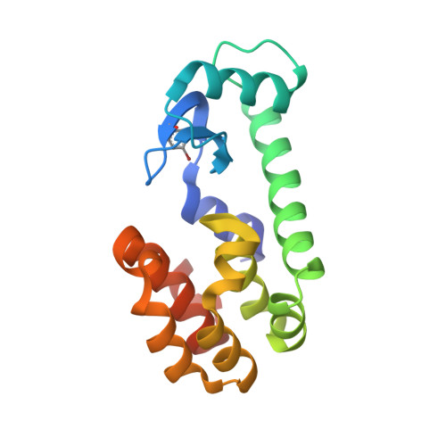
5KI3

PSEUDO T4 LYSOZYME MUTANT - Y18PHE-BR


Experimental Data Snapshot

  • Method: X-RAY DIFFRACTION
  • Resolution: 1.65 Å
  • R-Value Free: 0.250 
  • R-Value Work: 0.200 
  • R-Value Observed: 0.210 

Starting Model: experimental
View more details

wwPDB Validation   3D Report Full Report


Ligand Structure Quality Assessment 


This is version 1.6 of the entry. See complete history


Literature

Structure-Energy Relationships of Halogen Bonds in Proteins.

Scholfield, M.R.Ford, M.C.Carlsson, A.C.Butta, H.Mehl, R.A.Ho, P.S.

(2017) Biochemistry 56: 2794-2802

  • DOI: https://doi.org/10.1021/acs.biochem.7b00022
  • Primary Citation of Related Structures:  
    5KHZ, 5KI1, 5KI2, 5KI3, 5KI8, 5KIG, 5KII, 5KIM, 5KIO

  • PubMed Abstract: 

    The structures and stabilities of proteins are defined by a series of weak noncovalent electrostatic, van der Waals, and hydrogen bond (HB) interactions. In this study, we have designed and engineered halogen bonds (XBs) site-specifically to study their structure-energy relationship in a model protein, T4 lysozyme. The evidence for XBs is the displacement of the aromatic side chain toward an oxygen acceptor, at distances that are equal to or less than the sums of their respective van der Waals radii, when the hydroxyl substituent of the wild-type tyrosine is replaced by a halogen. In addition, thermal melting studies show that the iodine XB rescues the stabilization energy from an otherwise destabilizing substitution (at an equivalent noninteracting site), indicating that the interaction is also present in solution. Quantum chemical calculations show that the XB complements an HB at this site and that solvent structure must also be considered in trying to design molecular interactions such as XBs into biological systems. A bromine substitution also shows displacement of the side chain, but the distances and geometries do not indicate formation of an XB. Thus, we have dissected the contributions from various noncovalent interactions of halogens introduced into proteins, to drive the application of XBs, particularly in biomolecular design.


  • Organizational Affiliation

    Department of Biochemistry & Molecular Biology, Colorado State University , Fort Collins, Colorado 80523-1870, United States.


Macromolecules
Find similar proteins by:  (by identity cutoff)  |  3D Structure
Entity ID: 1
MoleculeChains Sequence LengthOrganismDetailsImage
Endolysin170Tequatrovirus T4Mutation(s): 2 
EC: 3.2.1.17
UniProt
Find proteins for P00720 (Enterobacteria phage T4)
Explore P00720 
Go to UniProtKB:  P00720
Entity Groups  
Sequence Clusters30% Identity50% Identity70% Identity90% Identity95% Identity100% Identity
UniProt GroupP00720
Sequence Annotations
Expand
  • Reference Sequence
Small Molecules
Ligands 1 Unique
IDChains Name / Formula / InChI Key2D Diagram3D Interactions
HED
Query on HED

Download Ideal Coordinates CCD File 
B [auth A]2-HYDROXYETHYL DISULFIDE
C4 H10 O2 S2
KYNFOMQIXZUKRK-UHFFFAOYSA-N
Modified Residues  1 Unique
IDChains TypeFormula2D DiagramParent
4BF
Query on 4BF
A
L-PEPTIDE LINKINGC9 H10 Br N O2TYR
Experimental Data & Validation

Experimental Data

  • Method: X-RAY DIFFRACTION
  • Resolution: 1.65 Å
  • R-Value Free: 0.250 
  • R-Value Work: 0.200 
  • R-Value Observed: 0.210 
  • Space Group: P 32 2 1
Unit Cell:
Length ( Å )Angle ( ˚ )
a = 59.589α = 90
b = 59.589β = 90
c = 95.15γ = 120
Software Package:
Software NamePurpose
d*TREKdata scaling
d*TREKdata reduction
PHASERphasing
PHENIXrefinement

Structure Validation

View Full Validation Report


Ligand Structure Quality Assessment 


Entry History & Funding Information

Deposition Data


Funding OrganizationLocationGrant Number
National Science Foundation (NSF, United States)United StatesCHE-1152494

Revision History  (Full details and data files)

  • Version 1.0: 2017-04-12
    Type: Initial release
  • Version 1.1: 2017-04-19
    Changes: Database references
  • Version 1.2: 2017-06-14
    Changes: Database references
  • Version 1.3: 2017-09-27
    Changes: Author supporting evidence
  • Version 1.4: 2019-11-27
    Changes: Author supporting evidence
  • Version 1.5: 2023-09-27
    Changes: Data collection, Database references, Refinement description
  • Version 1.6: 2023-11-15
    Changes: Data collection