5J21

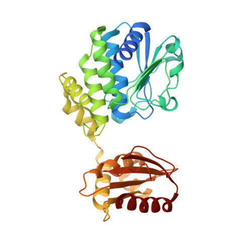
Structure of Bacillus NanoRNase A (WT)


Experimental Data Snapshot

  • Method: X-RAY DIFFRACTION
  • Resolution: 2.00 Å
  • R-Value Free: 0.232 
  • R-Value Work: 0.180 
  • R-Value Observed: 0.183 

Starting Model: experimental
View more details

wwPDB Validation   3D Report Full Report


This is version 1.4 of the entry. See complete history


Literature

Structural Basis for the Bidirectional Activity of Bacillus nanoRNase NrnA.

Schmier, B.J.Nelersa, C.M.Malhotra, A.

(2017) Sci Rep 7: 11085-11085

  • DOI: https://doi.org/10.1038/s41598-017-09403-x
  • Primary Citation of Related Structures:  
    5IPP, 5IUF, 5IZO, 5J21

  • PubMed Abstract: 

    NanoRNAs are RNA fragments 2 to 5 nucleotides in length that are generated as byproducts of RNA degradation and abortive transcription initiation. Cells have specialized enzymes to degrade nanoRNAs, such as the DHH phosphoesterase family member NanoRNase A (NrnA). This enzyme was originally identified as a 3' → 5' exonuclease, but we show here that NrnA is bidirectional, degrading 2-5 nucleotide long RNA oligomers from the 3' end, and longer RNA substrates from the 5' end. The crystal structure of Bacillus subtilis NrnA reveals a dynamic bi-lobal architecture, with the catalytic N-terminal DHH domain linked to the substrate binding C-terminal DHHA1 domain via an extended linker. Whereas this arrangement is similar to the structure of RecJ, a 5' → 3' DHH family DNase and other DHH family nanoRNases, Bacillus NrnA has gained an extended substrate-binding patch that we posit is responsible for its 3' → 5' activity.


  • Organizational Affiliation

    Department of Biochemistry and Molecular Biology, University of Miami Miller School of Medicine, PO Box 016129, Miami, FL, 33101-6129, USA.


Macromolecules
Find similar proteins by:  (by identity cutoff)  |  3D Structure
Entity ID: 1
MoleculeChains Sequence LengthOrganismDetailsImage
Bifunctional oligoribonuclease and PAP phosphatase NrnA
A, B, C, D
333Bacillus subtilis subsp. subtilis str. 168Mutation(s): 0 
Gene Names: nrnAytqIBSU29250
EC: 3.1.3.7 (PDB Primary Data), 3.1 (UniProt)
UniProt
Find proteins for O34600 (Bacillus subtilis (strain 168))
Explore O34600 
Go to UniProtKB:  O34600
Entity Groups  
Sequence Clusters30% Identity50% Identity70% Identity90% Identity95% Identity100% Identity
UniProt GroupO34600
Sequence Annotations
Expand
  • Reference Sequence
Experimental Data & Validation

Experimental Data

  • Method: X-RAY DIFFRACTION
  • Resolution: 2.00 Å
  • R-Value Free: 0.232 
  • R-Value Work: 0.180 
  • R-Value Observed: 0.183 
  • Space Group: P 1 21 1
Unit Cell:
Length ( Å )Angle ( ˚ )
a = 50.616α = 90
b = 121.25β = 91.31
c = 123.388γ = 90
Software Package:
Software NamePurpose
REFMACrefinement
HKL-2000data reduction
HKL-2000data scaling
PHASERphasing

Structure Validation

View Full Validation Report


Entry History & Funding Information

Deposition Data


Funding OrganizationLocationGrant Number
National Institutes of Health/National Institute of General Medical Sciences (NIH/NIGMS)United StatesGM69972

Revision History  (Full details and data files)

  • Version 1.0: 2017-08-02
    Type: Initial release
  • Version 1.1: 2017-09-13
    Changes: Author supporting evidence
  • Version 1.2: 2019-02-13
    Changes: Data collection, Database references
  • Version 1.3: 2019-12-25
    Changes: Author supporting evidence
  • Version 1.4: 2023-09-27
    Changes: Advisory, Data collection, Database references, Refinement description