5IQN

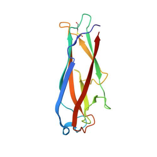
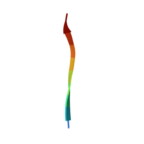
Crystal structure of the E. coli type 1 pilus subunit FimG (engineered variant with substitution Q134E; N-terminal FimG residues 1-12 truncated) in complex with the donor strand peptide DsF_SRIRIRGYVR


Experimental Data Snapshot

  • Method: X-RAY DIFFRACTION
  • Resolution: 1.00 Å
  • R-Value Free: 0.146 
  • R-Value Work: 0.118 
  • R-Value Observed: 0.119 

Starting Model: experimental
View more details

wwPDB Validation   3D Report Full Report


This is version 1.4 of the entry. See complete history


Literature

Accelerating the Association of the Most Stable Protein-Ligand Complex by More than Two Orders of Magnitude.

Giese, C.Eras, J.Kern, A.Scharer, M.A.Capitani, G.Glockshuber, R.

(2016) Angew Chem Int Ed Engl 55: 9350-9355

  • DOI: https://doi.org/10.1002/anie.201603652
  • Primary Citation of Related Structures:  
    5IQM, 5IQN, 5IQO

  • PubMed Abstract: 

    The complex between the bacterial type 1 pilus subunit FimG and the peptide corresponding to the N-terminal extension (termed donor strand, Ds) of the partner subunit FimF (DsF) shows the strongest reported noncovalent molecular interaction, with a dissociation constant (KD ) of 1.5×10(-20)  m. However, the complex only exhibits a slow association rate of 330 m(-1)  s(-1) that limits technical applications, such as its use in affinity purification. Herein, a structure-based approach was used to design pairs of FimGt (a FimG variant lacking its own N-terminal extension) and DsF variants with enhanced electrostatic surface complementarity. Association of the best mutant FimGt/DsF pairs was accelerated by more than two orders of magnitude, while the dissociation rates and 3D structures of the improved complexes remained essentially unperturbed. A KD  value of 8.8×10(-22)  m was obtained for the best mutant complex, which is the lowest value reported to date for a protein/ligand complex.


  • Organizational Affiliation

    Department of Biology, ETH Zurich, Otto-Stern-Weg 5, 8093, Zürich, Switzerland.


Macromolecules
Find similar proteins by:  (by identity cutoff)  |  3D Structure
Entity ID: 1
MoleculeChains Sequence LengthOrganismDetailsImage
Protein FimGA,
D [auth G]
132Escherichia coli K-12Mutation(s): 1 
Gene Names: fimGb4319JW4282
UniProt
Find proteins for P08190 (Escherichia coli (strain K12))
Explore P08190 
Go to UniProtKB:  P08190
Entity Groups  
Sequence Clusters30% Identity50% Identity70% Identity90% Identity95% Identity100% Identity
UniProt GroupP08190
Sequence Annotations
Expand
  • Reference Sequence

Find similar proteins by:  Sequence   |   3D Structure  

Entity ID: 2
MoleculeChains Sequence LengthOrganismDetailsImage
Protein FimFB,
C [auth F]
10Escherichia coli K-12Mutation(s): 2 
UniProt
Find proteins for P08189 (Escherichia coli (strain K12))
Explore P08189 
Go to UniProtKB:  P08189
Entity Groups  
Sequence Clusters30% Identity50% Identity70% Identity90% Identity95% Identity100% Identity
UniProt GroupP08189
Sequence Annotations
Expand
  • Reference Sequence
Experimental Data & Validation

Experimental Data

  • Method: X-RAY DIFFRACTION
  • Resolution: 1.00 Å
  • R-Value Free: 0.146 
  • R-Value Work: 0.118 
  • R-Value Observed: 0.119 
  • Space Group: P 1 21 1
Unit Cell:
Length ( Å )Angle ( ˚ )
a = 25.68α = 90
b = 52.88β = 98.71
c = 83.14γ = 90
Software Package:
Software NamePurpose
PHENIXrefinement
XDSdata reduction
XSCALEdata scaling
PHASERphasing

Structure Validation

View Full Validation Report


Entry History 

Deposition Data

Revision History  (Full details and data files)

  • Version 1.0: 2016-07-06
    Type: Initial release
  • Version 1.1: 2016-08-10
    Changes: Database references
  • Version 1.2: 2019-11-20
    Changes: Advisory, Data collection, Derived calculations
  • Version 1.3: 2024-01-10
    Changes: Advisory, Data collection, Database references, Derived calculations, Refinement description
  • Version 1.4: 2024-10-23
    Changes: Structure summary