5H42

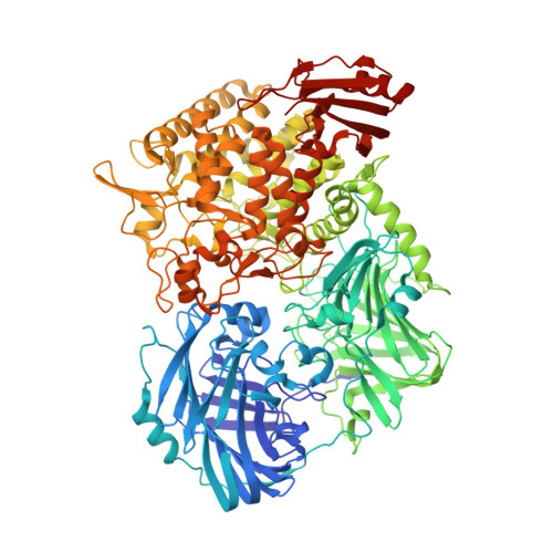
Crystal Structure of 1,2-beta-oligoglucan phosphorylase from Lachnoclostridium phytofermentans in complex with alpha-d-glucose-1-phosphate


Experimental Data Snapshot

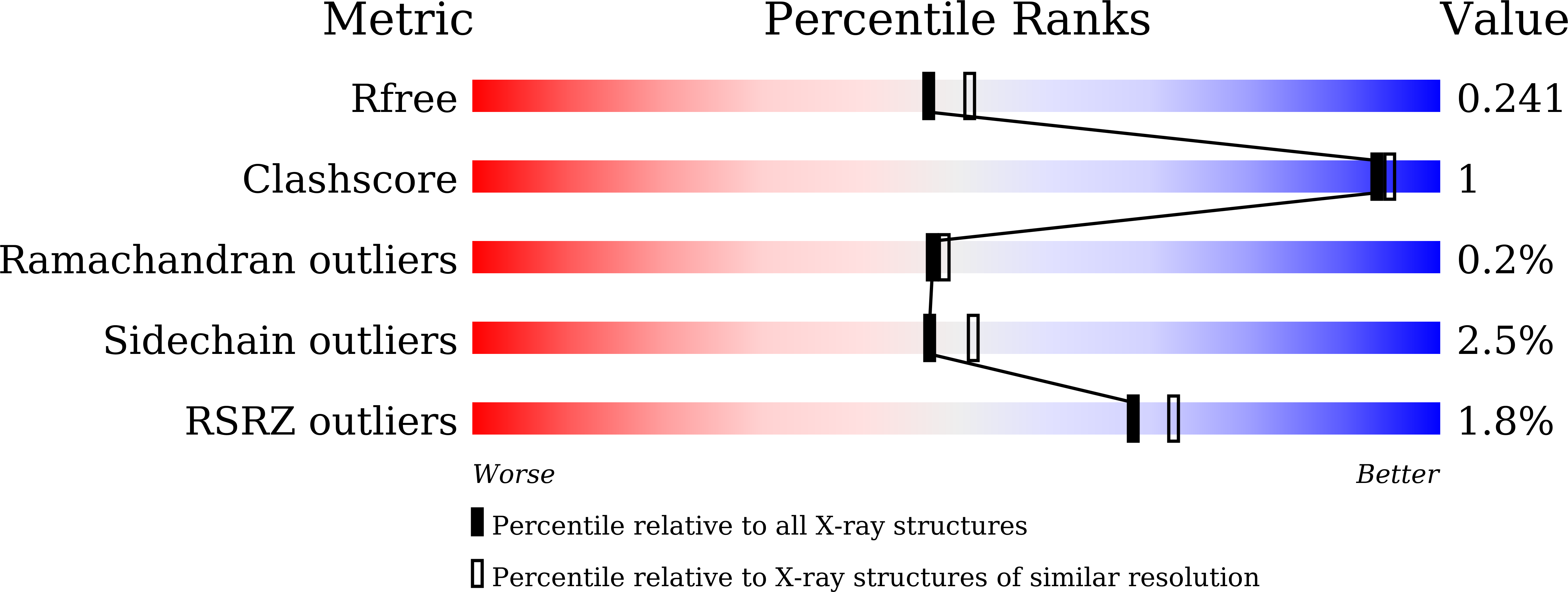
  • Method: X-RAY DIFFRACTION
  • Resolution: 2.10 Å
  • R-Value Free: 0.236 
  • R-Value Work: 0.195 
  • R-Value Observed: 0.197 

Starting Model: experimental
View more details

wwPDB Validation   3D Report Full Report


Ligand Structure Quality Assessment 


This is version 1.3 of the entry. See complete history


Literature

Mechanistic insight into the substrate specificity of 1,2-beta-oligoglucan phosphorylase from Lachnoclostridium phytofermentans

Nakajima, M.Tanaka, N.Furukawa, N.Nihira, T.Kodutsumi, Y.Takahashi, Y.Sugimoto, N.Miyanaga, A.Fushinobu, S.Taguchi, H.Nakai, H.

(2017) Sci Rep 7: 42671-42671

  • DOI: https://doi.org/10.1038/srep42671
  • Primary Citation of Related Structures:  
    5H3Z, 5H40, 5H41, 5H42

  • PubMed Abstract: 

    Glycoside phosphorylases catalyze the phosphorolysis of oligosaccharides into sugar phosphates. Recently, we found a novel phosphorylase acting on β-1,2-glucooligosaccharides with degrees of polymerization of 3 or more (1,2-β-oligoglucan phosphorylase, SOGP) in glycoside hydrolase family (GH) 94. Here, we characterized SOGP from Lachnoclostridium phytofermentans (LpSOGP) and determined its crystal structure. LpSOGP is a monomeric enzyme that contains a unique β-sandwich domain (Ndom1) at its N-terminus. Unlike the dimeric GH94 enzymes possessing catalytic pockets at their dimer interface, LpSOGP has a catalytic pocket between Ndom1 and the catalytic domain. In the complex structure of LpSOGP with sophorose, sophorose binds at subsites +1 to +2. Notably, the Glc moiety at subsite +1 is flipped compared with the corresponding ligands in other GH94 enzymes. This inversion suggests the great distortion of the glycosidic bond between subsites -1 and +1, which is likely unfavorable for substrate binding. Compensation for this disadvantage at subsite +2 can be accounted for by the small distortion of the glycosidic bond in the sophorose molecule. Therefore, the binding mode at subsites +1 and +2 defines the substrate specificity of LpSOGP, which provides mechanistic insights into the substrate specificity of a phosphorylase acting on β-1,2-glucooligosaccharides.


  • Organizational Affiliation

    Department of Applied Biological Science, Faculty of Science and Technology, Tokyo University of Science, Chiba, Japan.


Macromolecules
Find similar proteins by:  (by identity cutoff)  |  3D Structure
Entity ID: 1
MoleculeChains Sequence LengthOrganismDetailsImage
Uncharacterized protein
A, B
1,122Lachnoclostridium phytofermentans ISDgMutation(s): 0 
Gene Names: Cphy_0694
EC: 2.4.1.333
UniProt
Find proteins for A9KJS6 (Lachnoclostridium phytofermentans (strain ATCC 700394 / DSM 18823 / ISDg))
Explore A9KJS6 
Go to UniProtKB:  A9KJS6
Entity Groups  
Sequence Clusters30% Identity50% Identity70% Identity90% Identity95% Identity100% Identity
UniProt GroupA9KJS6
Sequence Annotations
Expand
  • Reference Sequence
Experimental Data & Validation

Experimental Data

  • Method: X-RAY DIFFRACTION
  • Resolution: 2.10 Å
  • R-Value Free: 0.236 
  • R-Value Work: 0.195 
  • R-Value Observed: 0.197 
  • Space Group: P 1 21 1
Unit Cell:
Length ( Å )Angle ( ˚ )
a = 86.469α = 90
b = 94.778β = 93.96
c = 156.851γ = 90
Software Package:
Software NamePurpose
REFMACrefinement
HKL-2000data reduction
HKL-2000data scaling
MOLREPphasing

Structure Validation

View Full Validation Report


Ligand Structure Quality Assessment 


Entry History 

Deposition Data

Revision History  (Full details and data files)

  • Version 1.0: 2017-03-01
    Type: Initial release
  • Version 1.1: 2020-02-26
    Changes: Data collection
  • Version 1.2: 2020-07-29
    Type: Remediation
    Reason: Carbohydrate remediation
    Changes: Data collection, Derived calculations, Structure summary
  • Version 1.3: 2023-11-08
    Changes: Data collection, Database references, Refinement description, Structure summary