Domain Movements upon Activation of Phenylalanine Hydroxylase Characterized by Crystallography and Chromatography-Coupled Small-Angle X-ray Scattering.
Meisburger, S.P., Taylor, A.B., Khan, C.A., Zhang, S., Fitzpatrick, P.F., Ando, N.(2016) J Am Chem Soc 138: 6506-6516
- PubMed: 27145334
- DOI: https://doi.org/10.1021/jacs.6b01563
- Primary Citation of Related Structures:
5EGQ, 5FGJ - PubMed Abstract:
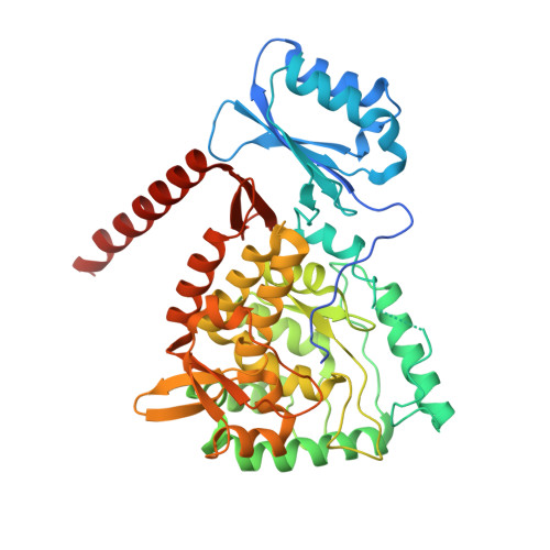
Mammalian phenylalanine hydroxylase (PheH) is an allosteric enzyme that catalyzes the first step in the catabolism of the amino acid phenylalanine. Following allosteric activation by high phenylalanine levels, the enzyme catalyzes the pterin-dependent conversion of phenylalanine to tyrosine. Inability to control elevated phenylalanine levels in the blood leads to increased risk of mental disabilities commonly associated with the inherited metabolic disorder, phenylketonuria. Although extensively studied, structural changes associated with allosteric activation in mammalian PheH have been elusive. Here, we examine the complex allosteric mechanisms of rat PheH using X-ray crystallography, isothermal titration calorimetry (ITC), and small-angle X-ray scattering (SAXS). We describe crystal structures of the preactivated state of the PheH tetramer depicting the regulatory domains docked against the catalytic domains and preventing substrate binding. Using SAXS, we further describe the domain movements involved in allosteric activation of PheH in solution and present the first demonstration of chromatography-coupled SAXS with Evolving Factor Analysis (EFA), a powerful method for separating scattering components in a model-independent way. Together, these results support a model for allostery in PheH in which phenylalanine stabilizes the dimerization of the regulatory domains and exposes the active site for substrate binding and other structural changes needed for activity.
Organizational Affiliation:
Department of Chemistry, Princeton University , Princeton, New Jersey 08544, United States.