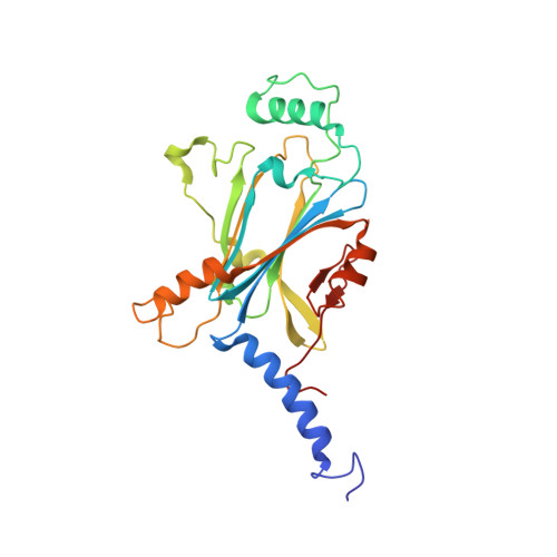
5EXY

Crystal structure of in cellulo recombinant CPV1 Polyhedra


Experimental Data Snapshot

  • Method: X-RAY DIFFRACTION
  • Resolution: 1.55 Å
  • R-Value Free: 0.167 
  • R-Value Work: 0.128 
  • R-Value Observed: 0.132 

Starting Model: experimental
View more details

wwPDB Validation   3D Report Full Report


Ligand Structure Quality Assessment 


This is version 1.2 of the entry. See complete history


Literature

A pipeline for structure determination of in vivo-grown crystals using in cellulo diffraction.

Boudes, M.Garriga, D.Fryga, A.Caradoc-Davies, T.Coulibaly, F.

(2016) Acta Crystallogr D Struct Biol 72: 576-585

  • DOI: https://doi.org/10.1107/S2059798316002369
  • Primary Citation of Related Structures:  
    5EXY, 5EXZ

  • PubMed Abstract: 

    While structure determination from micrometre-sized crystals used to represent a challenge, serial X-ray crystallography on microfocus beamlines at synchrotron and free-electron laser facilities greatly facilitates this process today for microcrystals and nanocrystals. In addition to typical microcrystals of purified recombinant protein, these advances have enabled the analysis of microcrystals produced inside living cells. Here, a pipeline where crystals are grown in insect cells, sorted by flow cytometry and directly analysed by X-ray diffraction is presented and applied to in vivo-grown crystals of the recombinant CPV1 polyhedrin. When compared with the analysis of purified crystals, in cellulo diffraction produces data of better quality and a gain of ∼0.35 Å in resolution for comparable beamtime usage. Importantly, crystals within cells are readily derivatized with gold and iodine compounds through the cellular membrane. Using the multiple isomorphous replacement method, a near-complete model was autobuilt from 2.7 Å resolution data. Thus, in favourable cases, an in cellulo pipeline can replace the complete workflow of structure determination without compromising the quality of the resulting model. In addition to its efficiency, this approach maintains the protein in a cellular context throughout the analysis, which reduces the risk of disrupting transient or labile interactions in protein-protein or protein-ligand complexes.


  • Organizational Affiliation

    Infection and Immunity Program, Monash Biomedicine Discovery Institute and Department of Biochemistry and Molecular Biology, Monash University, Melbourne, VIC 3800, Australia.


Macromolecules
Find similar proteins by:  (by identity cutoff)  |  3D Structure
Entity ID: 1
MoleculeChains Sequence LengthOrganismDetailsImage
Polyhedrin248Bombyx mori cypovirus 1Mutation(s): 1 
UniProt
Find proteins for P11041 (Bombyx mori cytoplasmic polyhedrosis virus)
Explore P11041 
Go to UniProtKB:  P11041
Entity Groups  
Sequence Clusters30% Identity50% Identity70% Identity90% Identity95% Identity100% Identity
UniProt GroupP11041
Sequence Annotations
Expand
  • Reference Sequence
Small Molecules
Ligands 5 Unique
IDChains Name / Formula / InChI Key2D Diagram3D Interactions
GTP
Query on GTP

Download Ideal Coordinates CCD File 
H [auth A]GUANOSINE-5'-TRIPHOSPHATE
C10 H16 N5 O14 P3
XKMLYUALXHKNFT-UUOKFMHZSA-N
ATP
Query on ATP

Download Ideal Coordinates CCD File 
F [auth A]ADENOSINE-5'-TRIPHOSPHATE
C10 H16 N5 O13 P3
ZKHQWZAMYRWXGA-KQYNXXCUSA-N
CTP
Query on CTP

Download Ideal Coordinates CCD File 
G [auth A]CYTIDINE-5'-TRIPHOSPHATE
C9 H16 N3 O14 P3
PCDQPRRSZKQHHS-XVFCMESISA-N
CL
Query on CL

Download Ideal Coordinates CCD File 
B [auth A]CHLORIDE ION
Cl
VEXZGXHMUGYJMC-UHFFFAOYSA-M
MG
Query on MG

Download Ideal Coordinates CCD File 
C [auth A],
D [auth A],
E [auth A]
MAGNESIUM ION
Mg
JLVVSXFLKOJNIY-UHFFFAOYSA-N
Experimental Data & Validation

Experimental Data

  • Method: X-RAY DIFFRACTION
  • Resolution: 1.55 Å
  • R-Value Free: 0.167 
  • R-Value Work: 0.128 
  • R-Value Observed: 0.132 
  • Space Group: I 2 3
Unit Cell:
Length ( Å )Angle ( ˚ )
a = 103.18α = 90
b = 103.18β = 90
c = 103.18γ = 90
Software Package:
Software NamePurpose
DENZOdata reduction
SCALEPACKdata scaling
Cootmodel building
PDB_EXTRACTdata extraction
BUSTERrefinement
BUSTERphasing

Structure Validation

View Full Validation Report


Ligand Structure Quality Assessment 


Entry History 

Deposition Data

Revision History  (Full details and data files)

  • Version 1.0: 2016-05-25
    Type: Initial release
  • Version 1.1: 2023-09-27
    Changes: Data collection, Database references, Derived calculations, Refinement description
  • Version 1.2: 2024-11-06
    Changes: Structure summary