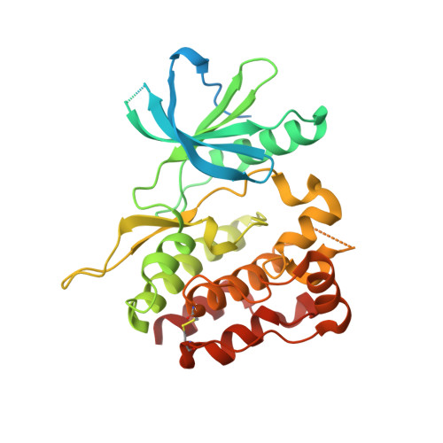
Structural and Functional Characterization of the JH2 Pseudokinase Domain of JAK Family Tyrosine Kinase 2 (TYK2).
Min, X., Ungureanu, D., Maxwell, S., Hammaren, H., Thibault, S., Hillert, E.K., Ayres, M., Greenfield, B., Eksterowicz, J., Gabel, C., Walker, N., Silvennoinen, O., Wang, Z.(2015) J Biol Chem 290: 27261-27270
- PubMed: 26359499
- DOI: https://doi.org/10.1074/jbc.M115.672048
- Primary Citation of Related Structures:
5C01, 5C03 - PubMed Abstract:
JAK (Janus family of cytoplasmic tyrosine kinases) family tyrosine kinase 2 (TYK2) participates in signaling through cytokine receptors involved in immune responses and inflammation. JAKs are characterized by dual kinase domain: a tyrosine kinase domain (JH1) that is preceded by a pseudokinase domain (JH2). The majority of disease-associated mutations in JAKs map to JH2, demonstrating its central regulatory function. JH2s were considered catalytically inactive, but JAK2 JH2 was found to have low autoregulatory catalytic activity. Whether the other JAK JH2s share ATP binding and enzymatic activity has been unclear. Here we report the crystal structure of TYK2 JH2 in complex with adenosine 5'-O-(thiotriphosphate) (ATP-γS) and characterize its nucleotide binding by biochemical and biophysical methods. TYK2 JH2 did not show phosphotransfer activity, but it binds ATP and the nucleotide binding stabilizes the protein without inducing major conformational changes. Mutation of the JH2 ATP-binding pocket increased basal TYK2 phosphorylation and downstream signaling. The overall structural characteristics of TYK2 JH2 resemble JAK2 JH2, but distinct stabilizing molecular interactions around helix αAL in the activation loop provide a structural basis for differences in substrate access and catalytic activities among JAK family JH2s. The structural and biochemical data suggest that ATP binding is functionally important for both TYK2 and JAK2 JH2s, whereas the regulatory phosphorylation appears to be a unique property of JAK2. Finally, the co-crystal structure of TYK2 JH2 complexed with a small molecule inhibitor demonstrates that JH2 is accessible to ATP-competitive compounds, which offers novel approaches for targeting cytokine signaling as well as potential therapeutic applications.
Organizational Affiliation:
Departments of Therapeutic Discovery, Amgen Inc., South San Francisco, California 94080.


