5BS3

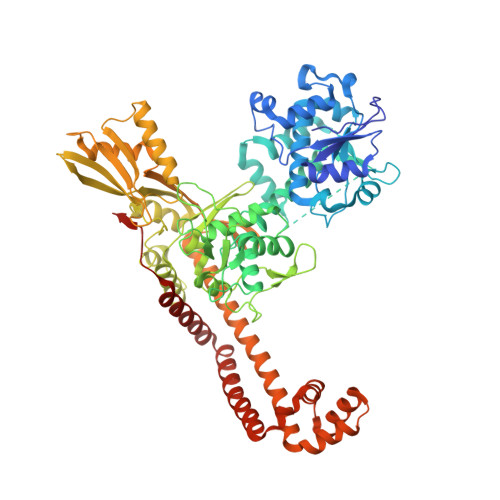
Crystal Structure of S.A. gyrase in complex with Compound 7


Experimental Data Snapshot

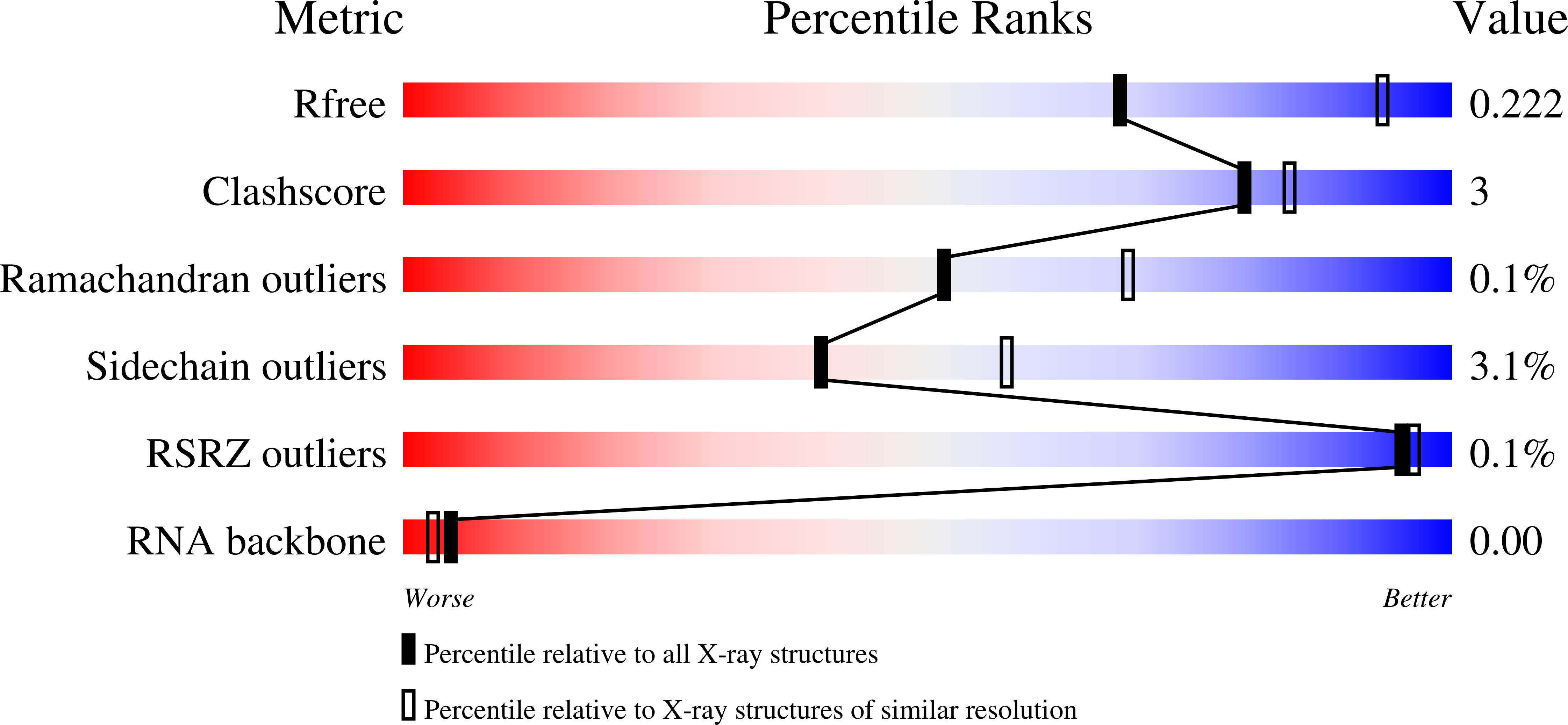
  • Method: X-RAY DIFFRACTION
  • Resolution: 2.65 Å
  • R-Value Free: 0.213 
  • R-Value Work: 0.170 
  • R-Value Observed: 0.172 

wwPDB Validation   3D Report Full Report


Ligand Structure Quality Assessment 


This is version 1.1 of the entry. See complete history


Literature

Tricyclic 1,5-naphthyridinone oxabicyclooctane-linked novel bacterial topoisomerase inhibitors as broad-spectrum antibacterial agents-SAR of left-hand-side moiety (Part-2).

Singh, S.B.Kaelin, D.E.Wu, J.Miesel, L.Tan, C.M.Black, T.Nargund, R.Meinke, P.T.Olsen, D.B.Lagrutta, A.Lu, J.Patel, S.Rickert, K.W.Smith, R.F.Soisson, S.Sherer, E.Joyce, L.A.Wei, C.Peng, X.Wang, X.Fukuda, H.Kishii, R.Takei, M.Takano, H.Shibasaki, M.Yajima, M.Nishimura, A.Shibata, T.Fukuda, Y.

(2015) Bioorg Med Chem Lett 25: 1831-1835

  • DOI: https://doi.org/10.1016/j.bmcl.2015.03.044
  • Primary Citation of Related Structures:  
    5BS3

  • PubMed Abstract: 

    Novel bacterial topoisomerase inhibitors (NBTIs) represent a new class of broad-spectrum antibacterial agents targeting bacterial Gyrase A and ParC and have potential utility in combating antibiotic resistance. A series of novel oxabicyclooctane-linked NBTIs with new tricyclic-1,5-naphthyridinone left hand side moieties have been described. Compounds with a (R)-hydroxy-1,5-naphthyridinone moiety (7) showed potent antibacterial activity (e.g., Staphylococcus aureus MIC 0.25 μg/mL), acceptable Gram-positive and Gram-negative spectrum with rapidly bactericidal activity. The compound 7 showed intravenous and oral efficacy (ED50) at 3.2 and 27 mg/kg doses, respectively, in a murine model of bacteremia. Most importantly they showed significant attenuation of functional hERG activity (IC50 >170 μM). In general, lower logD attenuated hERG activity but also reduced Gram-negative activity. The co-crystal structure of a hydroxy-tricyclic NBTI bound to a DNA-gyrase complex exhibited a binding mode that show enantiomeric preference for R isomer and explains the activity and SAR. The discovery, synthesis, SAR and X-ray crystal structure of the left-hand-side tricyclic 1,5-naphthyridinone based oxabicyclooctane linked NBTIs are described.


  • Organizational Affiliation

    Merck Research Laboratories, Kenilworth, NJ 07033, United States. Electronic address: sheo.singh.215@gmail.com.


Macromolecules

Find similar proteins by:  (by identity cutoff)  |  3D Structure
Entity ID: 1
MoleculeChains Sequence LengthOrganismDetailsImage
DNA gyrase subunit A and BA [auth B],
B [auth D]
692Staphylococcus aureusMutation(s): 2 
Gene Names: gyrBgyrA
EC: 5.99.1.3 (PDB Primary Data), 5.6.2.2 (UniProt)
UniProt
Find proteins for P20831 (Staphylococcus aureus)
Explore P20831 
Go to UniProtKB:  P20831
Find proteins for P0A0K8 (Staphylococcus aureus)
Explore P0A0K8 
Go to UniProtKB:  P0A0K8
Entity Groups  
Sequence Clusters30% Identity50% Identity70% Identity90% Identity95% Identity100% Identity
UniProt GroupsP20831P0A0K8
Sequence Annotations
Expand
  • Reference Sequence

Find similar nucleic acids by:  Sequence   |   3D Structure  

Entity ID: 2
MoleculeChains LengthOrganismImage
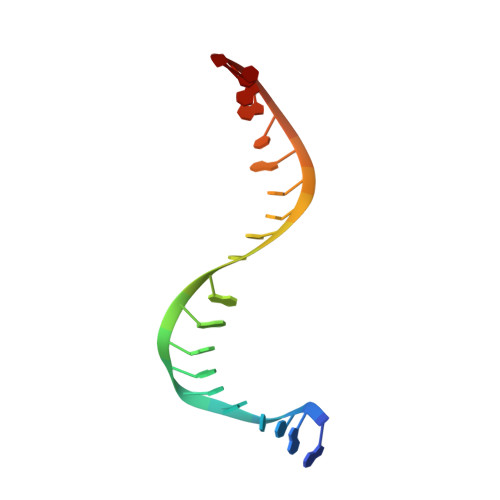
DNA/RNA (5'-R(P*AP*GP*CP*CP*G)-D(P*T)-R(P*AP*GP*GP*GP*CP*CP*C)-D(P*T)-R(P*AP*CP*GP*GP*C)-D(P*T)-3')C [auth E],
D [auth F]
20Staphylococcus aureus subsp. aureus ED98
Sequence Annotations
Expand
  • Reference Sequence
Small Molecules
Ligands 3 Unique
IDChains Name / Formula / InChI Key2D Diagram3D Interactions
WCP
Query on WCP

Download Ideal Coordinates CCD File 
I [auth F](4R)-3-fluoro-4-hydroxy-4-{[(1r,4R)-4-{[(3-oxo-3,4-dihydro-2H-pyrido[3,2-b][1,4]oxazin-6-yl)methyl]amino}-2-oxabicyclo[2.2.2]oct-1-yl]methyl}-4,5-dihydro-7H-pyrrolo[3,2,1-de][1,5]naphthyridin-7-one
C26 H26 F N5 O5
XTHQMKOHGLBSON-GSDHBNRESA-N
SO4
Query on SO4

Download Ideal Coordinates CCD File 
G [auth D],
H [auth E],
J [auth F]
SULFATE ION
O4 S
QAOWNCQODCNURD-UHFFFAOYSA-L
MN
Query on MN

Download Ideal Coordinates CCD File 
E [auth B],
F [auth D]
MANGANESE (II) ION
Mn
WAEMQWOKJMHJLA-UHFFFAOYSA-N
Experimental Data & Validation

Experimental Data

  • Method: X-RAY DIFFRACTION
  • Resolution: 2.65 Å
  • R-Value Free: 0.213 
  • R-Value Work: 0.170 
  • R-Value Observed: 0.172 
  • Space Group: P 61
Unit Cell:
Length ( Å )Angle ( ˚ )
a = 93.337α = 90
b = 93.337β = 90
c = 411.068γ = 120
Software Package:
Software NamePurpose
BUSTERrefinement
XDSdata reduction
SCALAdata scaling
REFMACphasing

Structure Validation

View Full Validation Report


Ligand Structure Quality Assessment 


Entry History 

Deposition Data

Revision History  (Full details and data files)

  • Version 1.0: 2015-06-17
    Type: Initial release
  • Version 1.1: 2024-03-06
    Changes: Data collection, Database references, Derived calculations, Source and taxonomy, Structure summary